
Professor of Medicine, University of Zaragoza
Dr. Borja del Carmelo Gracia Tello is an internist and academic director of the Unidad de Enfermedades Autoinmunes Sistémicas (Systemic Autoimmune Diseases Unit) at Hospital Clínico Universitario Lozano Blesa (Zaragoza), and Professor of Medicine at the University of Zaragoza. After completing his specialist training in Internal Medicine, he undertook a research fellowship with Prof. David Isenberg at University College London, where he gained advanced expertise in systemic autoimmune diseases and lupus. His clinical and research work focuses on microvascular involvement and connective tissue diseases, with a particular interest in systemic sclerosis, biomarkers of disease activity, and imaging-based tools to improve early diagnosis and risk stratification.
Dr. Gracia Tello has coordinated and participated in multiple national and international research projects, including studies on systemic sclerosis–associated pulmonary arterial hypertension, sarcoidosis, giant cell arteritis, and COVID-19. He is the lead inventor of a project on the digitalisation and automated analysis of nailfold capillaroscopy, developed through the GEAS (Grupo Español de Enfermedades Autoinmunes Sistémicas) of SEMI (Spanish Society of Internal Medicine), and co-author of the CAPI-Score and CAPI-Detect algorithms for quantitative videocapillaroscopy. As Chair of the Scientific Advisory Board, he provides strategic and scientific oversight for Capillaroscopy.com, ensuring that all educational content is rigorous, up-to-date, and aligned with international standards of care.
Contributed cases and studies (2)
Early SSc Cutolo Pattern: Representative Case
Complete capillaroscopy study illustrating early scleroderma-pattern microangiopathy with preserved density.
Active SSc Cutolo Pattern: Representative Case
Complete capillaroscopy study with prominent giant capillaries, hemorrhages, and moderate capillary loss.
Guides with credited review
Images in the atlas (202)
Showing 157–168 of 202 images
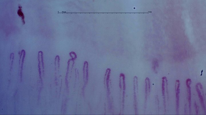
Mostly simple hairpin capillaries with one small coiled apex. An isolated elongated hemorrhage lies above the left side.
Recorded diagnosis: Systemic lupus erythematosus
- Magnification:
- 250×
- Device:
- Smart G-Scope
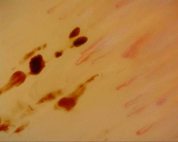
Fine elongated capillaries with numerous brown-red hemorrhagic deposits of different sizes.
- Magnification:
- 207.55×
- Device:
- Dino-Lite Edge
- Field of view (W × H):
- ≈ 1.88 × 1.50 mm
For the subset of apices with stable automated measurements, estimated widths span approximately 17–37 µm. These are automated estimates, not manually validated measurements.
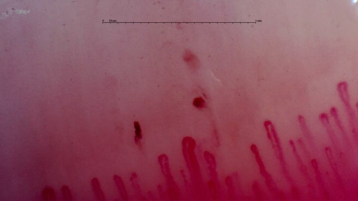
Capillaries with enlarged rounded apices and three separate reddish hemorrhagic deposits above the row.
Recorded diagnosis: Raynaud phenomenon
- Magnification:
- 250×
- Device:
- Smart G-Scope
- Field of view (W × H):
- ≈ 2.33 × 1.31 mm
For the subset of apices with stable automated measurements, estimated widths span approximately 23–45 µm. These are automated estimates, not manually validated measurements.
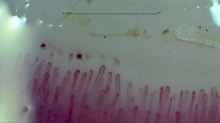
Elongated hairpin capillaries of different widths, with several small microhemorrhages above the left half.
Recorded diagnosis: Systemic lupus erythematosus
- Magnification:
- 250×
- Device:
- Smart G-Scope
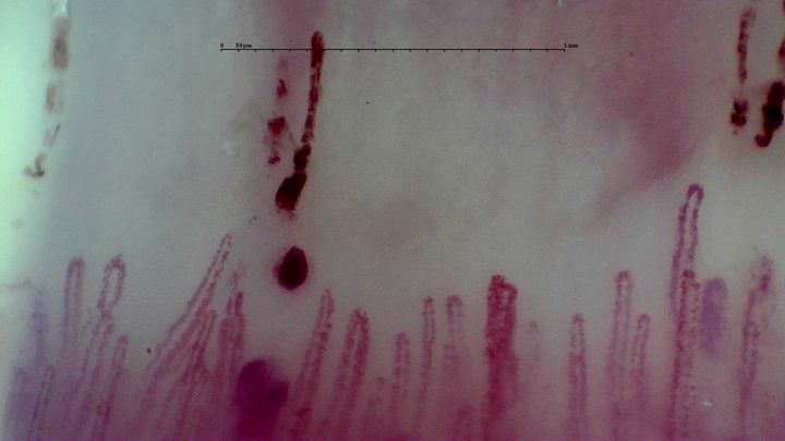
Broad capillaries with elongated and rounded hemorrhagic deposits above the center and right side.
- Magnification:
- 250×
- Device:
- Smart G-Scope
- Field of view (W × H):
- ≈ 2.29 × 1.29 mm
For the subset of apices with stable automated measurements, estimated widths span approximately 15–47 µm. These are automated estimates, not manually validated measurements.
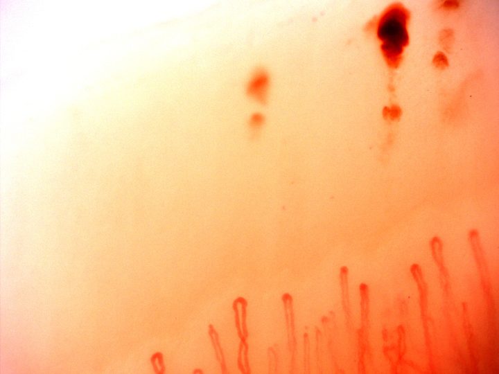
Slender capillary loops with a large dark red hemorrhagic deposit and smaller nearby foci at the upper right.
- Magnification:
- 200×
- Device:
- Optilia Digital Capillaroscope
- Field of view (W × H):
- ≈ 1.71 × 1.28 mm
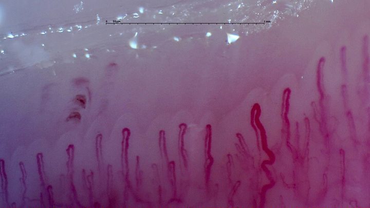
A long tortuous capillary on the right, beside straighter loops. Several small hemorrhagic deposits lie above the left half.
Recorded diagnosis: Raynaud phenomenon
- Magnification:
- 250×
- Device:
- Smart G-Scope
- Field of view (W × H):
- ≈ 2.32 × 1.31 mm
For the subset of apices with stable automated measurements, estimated widths span approximately 14–42 µm. These are automated estimates, not manually validated measurements.
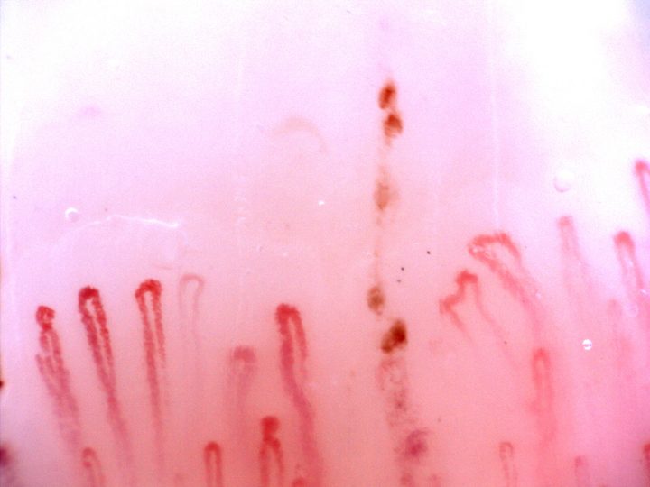
Capillaries with rounded apices of different widths and some irregular limbs. Brown-red hemorrhagic deposits form a vertical row near the center-right.
- Magnification:
- 200×
- Device:
- Optilia
- Field of view (W × H):
- ≈ 1.71 × 1.28 mm
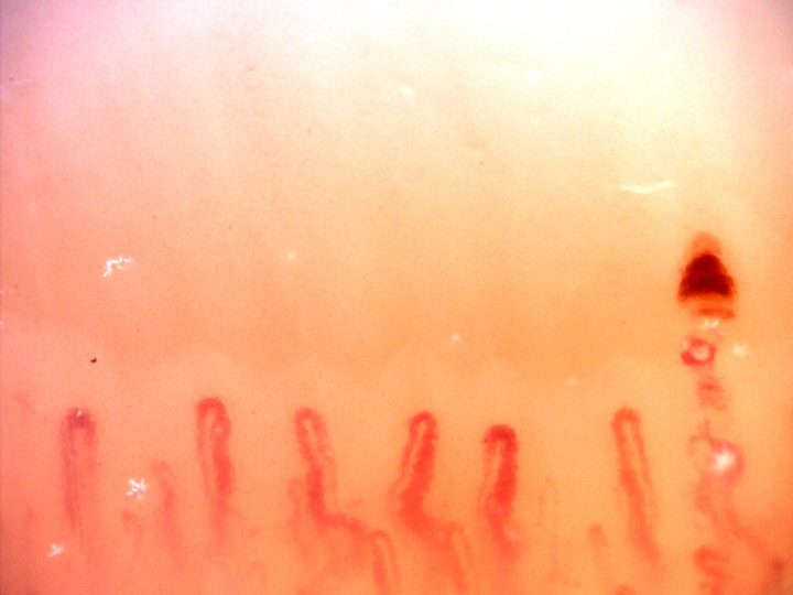
Rounded capillary loops with some curved limbs and a dense dark hemorrhagic deposit above the right side.
- Magnification:
- 200×
- Device:
- Optilia Digital Capillaroscope
- Field of view (W × H):
- ≈ 1.71 × 1.28 mm
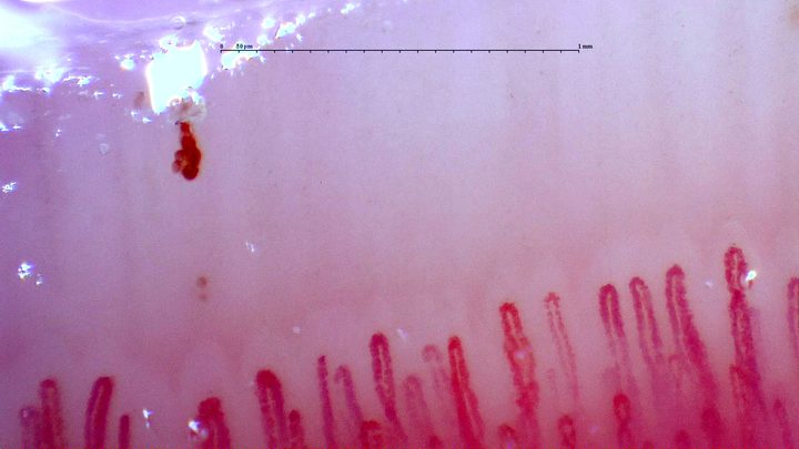
Fine and markedly enlarged capillaries, some partly obscured. An elongated dark hemorrhagic deposit lies above the left side.
- Magnification:
- 250×
- Device:
- Smart G-Scope
- Field of view (W × H):
- ≈ 2.24 × 1.26 mm
For the subset of apices with stable automated measurements, estimated widths span approximately 14–57 µm. These are automated estimates, not manually validated measurements.
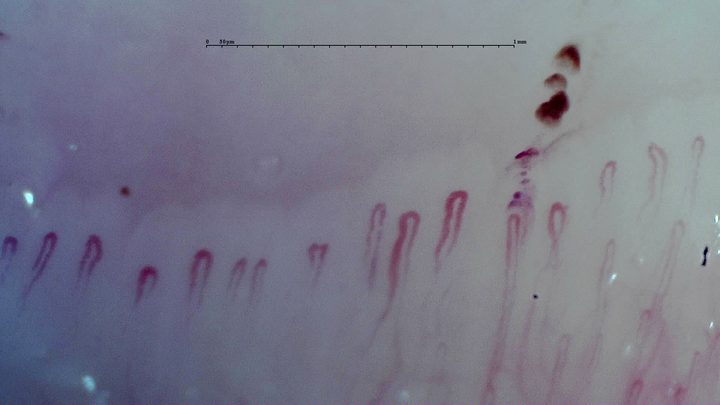
Mostly simple hairpin capillaries with rounded apices. Several dark hemorrhagic deposits lie above the center-right.
- Magnification:
- 250×
- Device:
- Smart G-Scope
- Field of view (W × H):
- ≈ 2.35 × 1.32 mm
For the subset of apices with stable automated measurements, estimated widths span approximately 18–30 µm. These are automated estimates, not manually validated measurements.
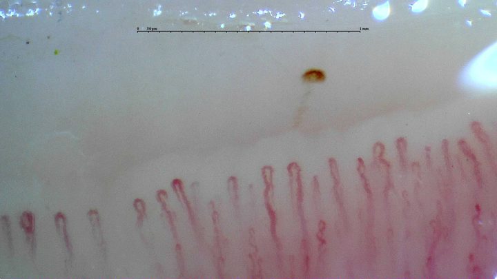
Fine hairpin capillaries with mild tortuosity and an isolated brown deposit compatible with a microhemorrhage above the row.
- Magnification:
- 250×
- Device:
- Smart G-Scope