Image Library

Searchable collection of capillaroscopic images organized by pattern, diagnosis, and severity.

Selected examples

Start exploring

Compare capillary shapes, sizes and hemorrhages.

Browse all images (243) →
200x capillaroscopy field in systemic sclerosis showing very dilated megacapillaries
View details

Large megacapillary (field 3)

Author: Miguel Antonio Mesa Navas

Magnification:
200×
Device:
Inspectis Digital Capillaroscope
Field of view (W × H):
≈ 1.68 × 1.26 mm
Systemic sclerosisGiant capillaryModerate to Severe
200x capillaroscopy field in systemic sclerosis showing a representative microvascular hemorrhagic lesion
View details

Hemorrhagic field in systemic sclerosis

Author: Miguel Antonio Mesa Navas

Magnification:
200×
Device:
Inspectis Digital Capillaroscope
Field of view (W × H):
≈ 1.68 × 1.26 mm
Systemic sclerosisHemorrhagesModerate to Severe
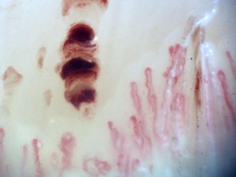
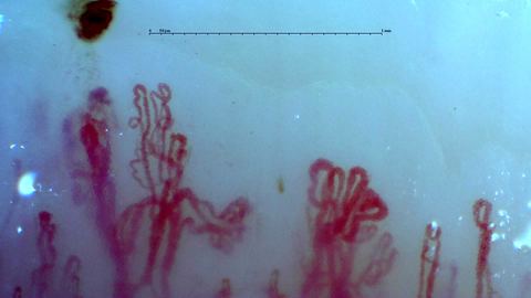
Two branching capillaries with multiple rounded apices and lateral branches. A dense dark hemorrhagic deposit lies above the left capillary.
View details

Two branching capillaries beneath a dark deposit

Author: Borja Gracia Tello

Magnification:
250×
Device:
Smart G-Scope
Field of view (W × H):
≈ 2.30 × 1.29 mm
Abnormal shapes and ramifications
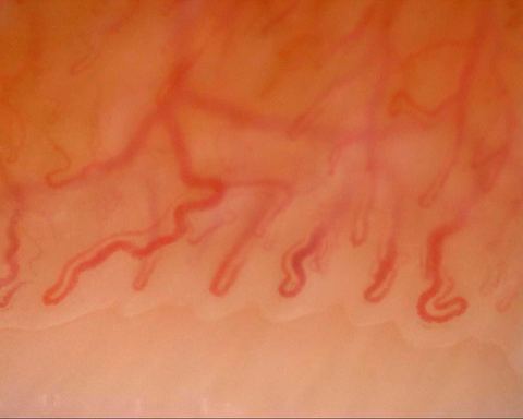
A long tortuous capillary on the left and a folded loop at the lower right, above a deeper transverse vessel.
View details

Broad tortuous loops above a transverse vessel

Author: Borja Gracia Tello

Magnification:
216.48×
Device:
Dino-Lite Edge
Field of view (W × H):
≈ 1.81 × 1.45 mm
Tortuous capillaries
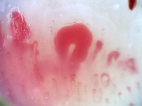
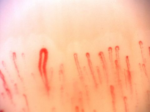
An enlarged capillary with a simple hairpin shape left of center, surrounded by finer loops.
View details

One broad open hairpin among fine capillaries

Author: Borja Gracia Tello

Magnification:
200×
Device:
Optilia Digital Capillaroscope
Field of view (W × H):
≈ 1.71 × 1.28 mm
Capillary dilation
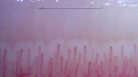
Narrow hairpin capillaries with small differences in length and width.
View details

Narrow rounded apices along a curved row

Author: Borja Gracia Tello

Magnification:
250×
Device:
Smart G-Scope
Field of view (W × H):
≈ 2.24 × 1.26 mm
Antiphospholipid syndromeSystemic lupus erythematosusSjögren syndromeSimple hairpin loops
200x nailfold capillaroscopy field with several megacapillaries in an active scleroderma pattern
View details

Active scleroderma pattern with megacapillaries (field 1)

Author: Miguel Antonio Mesa Navas

Magnification:
200×
Device:
Inspectis Digital Capillaroscope
Field of view (W × H):
≈ 1.68 × 1.26 mm
Systemic sclerosisActive scleroderma patternModerate to Severe
200x nailfold capillaroscopy field with prominent megacapillaries in an active scleroderma pattern
View details

Active scleroderma pattern with megacapillaries (field 2)

Author: Miguel Antonio Mesa Navas

Magnification:
200×
Device:
Inspectis Digital Capillaroscope
Field of view (W × H):
≈ 1.68 × 1.26 mm
Systemic sclerosisActive scleroderma patternModerate to Severe
200x nailfold capillaroscopy field with visible megacapillaries in an active scleroderma pattern
View details

Active scleroderma pattern with megacapillaries (field 3)

Author: Miguel Antonio Mesa Navas

Magnification:
200×
Device:
Inspectis Digital Capillaroscope
Field of view (W × H):
≈ 1.68 × 1.26 mm
Systemic sclerosisActive scleroderma patternModerate to Severe
200x nailfold capillaroscopy field with large megacapillaries in an active scleroderma pattern
View details

Active scleroderma pattern with megacapillaries (field 4)

Author: Miguel Antonio Mesa Navas

Magnification:
200×
Device:
Inspectis Digital Capillaroscope
Field of view (W × H):
≈ 1.68 × 1.26 mm
Systemic sclerosisActive scleroderma patternModerate to Severe
200x nailfold capillaroscopy field with a branching capillary on the left side
View details

Branching capillary on the left side

Author: Miguel Antonio Mesa Navas

Magnification:
200×
Device:
Inspectis Digital Capillaroscope
Field of view (W × H):
≈ 1.68 × 1.26 mm
Branching capillary / neoangiogenesis
200x capillaroscopy field with disappearance of the capillary bed on the left margin due to excessive pressure on the nail
View details

Pressure artifact with loss of visible capillary bed

Author: Miguel Antonio Mesa Navas

Magnification:
200×
Device:
Inspectis Digital Capillaroscope
Field of view (W × H):
≈ 1.68 × 1.26 mm
Acquisition artifactPressure artifact
200x capillaroscopy field showing a broad capillary-free space compatible with an avascular area
View details

Extensive avascular area

Author: Miguel Antonio Mesa Navas

Magnification:
200×
Device:
Inspectis Digital Capillaroscope
Field of view (W × H):
≈ 1.68 × 1.26 mm
Extensive avascular areaSevere
200x capillaroscopy field from systemic sclerosis showing marked architectural disorganization and neoangiogenesis without megacapillaries
View details

Late scleroderma pattern with severe disorganization (field 1)

Author: Miguel Antonio Mesa Navas

Magnification:
200×
Device:
Inspectis Digital Capillaroscope
Field of view (W × H):
≈ 1.68 × 1.26 mm
Systemic sclerosisLate scleroderma patternSevere
200x capillaroscopy field from systemic sclerosis showing severe architectural disorganization and neoangiogenesis without megacapillaries
View details

Late scleroderma pattern with severe disorganization (field 2)

Author: Miguel Antonio Mesa Navas

Magnification:
200×
Device:
Inspectis Digital Capillaroscope
Field of view (W × H):
≈ 1.68 × 1.26 mm
Systemic sclerosisLate scleroderma patternSevere
200x capillaroscopy field with a fog-like pericapillary haze between capillaries consistent with pericapillary edema
View details

Pericapillary edema on an active background

Author: Miguel Antonio Mesa Navas

Magnification:
200×
Device:
Inspectis Digital Capillaroscope
Field of view (W × H):
≈ 1.68 × 1.26 mm
Systemic sclerosisActive scleroderma patternModerate to Severe
200x capillaroscopy field in systemic sclerosis showing megacapillaries with a ground-glass stromal appearance suggestive of fibrosis
View details

Ground-glass stromal fibrosis with megacapillaries (field 1)

Author: Miguel Antonio Mesa Navas

Magnification:
200×
Device:
Inspectis Digital Capillaroscope
Field of view (W × H):
≈ 1.68 × 1.26 mm
Systemic sclerosisActive scleroderma pattern with stromal fibrosisSevere
200x capillaroscopy field in systemic sclerosis showing megacapillaries, pericapillary edema, and a ground-glass stromal pattern
View details

Ground-glass stromal fibrosis with megacapillaries (field 2)

Author: Miguel Antonio Mesa Navas

Magnification:
200×
Device:
Inspectis Digital Capillaroscope
Field of view (W × H):
≈ 1.68 × 1.26 mm
Systemic sclerosisActive scleroderma pattern with stromal fibrosisSevere
200x capillaroscopy field in systemic sclerosis showing a hemorrhage with a smoke-plume appearance
View details

Hemorrhage with smoke-plume appearance

Author: Miguel Antonio Mesa Navas

Magnification:
200×
Device:
Inspectis Digital Capillaroscope
Field of view (W × H):
≈ 1.68 × 1.26 mm
Systemic sclerosisHemorrhagesModerate to Severe
200x capillaroscopy field capturing a hemorrhage at the moment of its formation
View details

In situ hemorrhage formation

Author: Miguel Antonio Mesa Navas

Magnification:
200×
Device:
Inspectis Digital Capillaroscope
Field of view (W × H):
≈ 1.68 × 1.26 mm
Systemic sclerosisHemorrhagesModerate to Severe
200x capillaroscopy field in systemic sclerosis showing a very large megacapillary
View details

Large megacapillary (field 1)

Author: Miguel Antonio Mesa Navas

Magnification:
200×
Device:
Inspectis Digital Capillaroscope
Field of view (W × H):
≈ 1.68 × 1.26 mm
Systemic sclerosisGiant capillaryModerate to Severe
200x capillaroscopy field in systemic sclerosis showing a prominent megacapillary
View details

Large megacapillary (field 2)

Author: Miguel Antonio Mesa Navas

Magnification:
200×
Device:
Inspectis Digital Capillaroscope
Field of view (W × H):
≈ 1.68 × 1.26 mm
Systemic sclerosisGiant capillaryModerate to Severe
200x capillaroscopy field showing a giant capillary with a coiled appearance
View details

Coiled giant capillary

Author: Miguel Antonio Mesa Navas

Magnification:
200×
Device:
Inspectis Digital Capillaroscope
Field of view (W × H):
≈ 1.68 × 1.26 mm
Systemic sclerosisCoiled giant capillaryModerate to Severe
Capillaroscopy field from a critically ischemic digit with diffuse capillary thrombosis
View details

Suspected capillary thrombosis in critical digital ischemia

Author: Miguel Antonio Mesa Navas

Magnification:
200×
Device:
Inspectis Digital Capillaroscope
Field of view (W × H):
≈ 1.68 × 1.26 mm
Critical digital ischemiaSuspected capillary thrombosisSevere

Image Usage Guidelines

The atlas is an educational resource. Reuse rights are specific to each image: consult its detail page and the Image licensing and reuse page linked in the footer.

Images explicitly marked CC BY 4.0 may be reused, adapted and published, including commercially, under that licence without additional permission. Other images retain their individual terms.

Consent for educational publication does not by itself establish consent for broader reuse. A copyright licence does not settle patient privacy or other applicable rights.