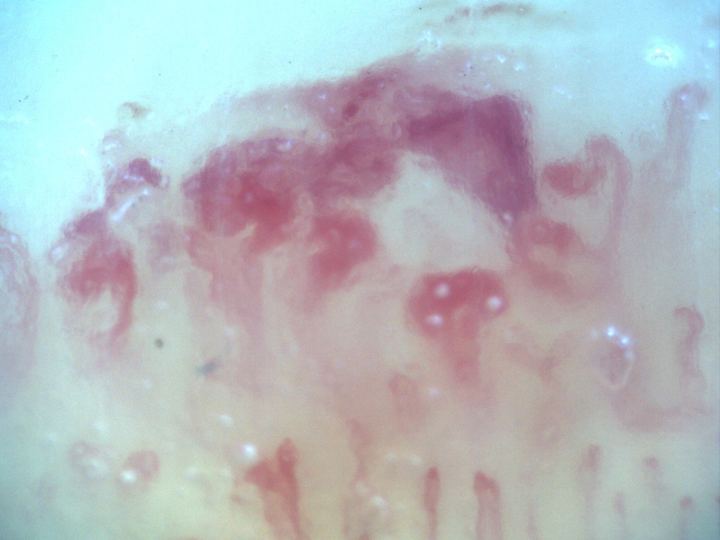
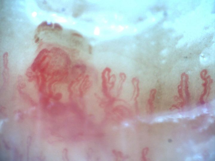
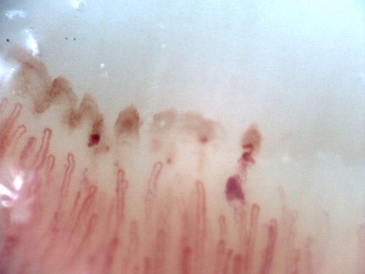
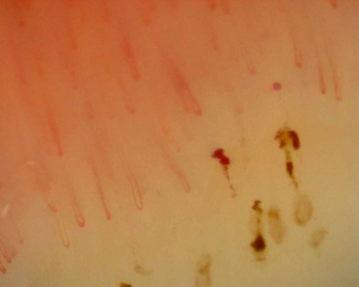
Hemorrhagic field in systemic sclerosis
200x capillaroscopy image from systemic sclerosis showing a representative microvascular hemorrhagic lesion.
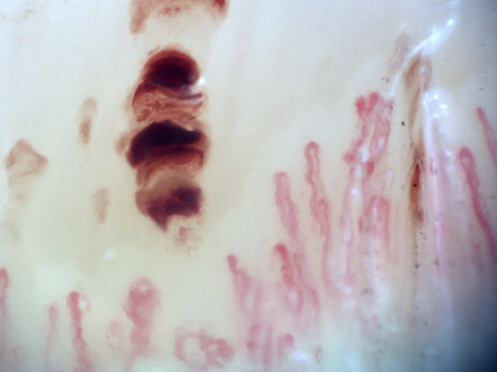
Image details
- Pathology
- Systemic sclerosis
- Pattern
- Hemorrhages
- Severity
- Moderate to Severe
- Author or contributor
- Miguel Antonio Mesa Navas
- Hallmarks
- Hemorrhages
- Magnification
- 200×
- Device
- Inspectis Digital Capillaroscope
- Field of view (W × H)
- ≈ 1.68 × 1.26 mm
Teaching point
Local trauma, manicure, and clinical context should be reviewed.
Clinical caveat
This image illustrates a visual finding and should be interpreted with acquisition quality, distribution across fingers, clinical context, and serology when relevant.
Use and licensing
Contributed by Miguel Antonio Mesa Navas for educational publication on Capillaroscopy.com. For reuse permissions, contact the rights holder.