Juvenile dermatomyositis: nailfold overview
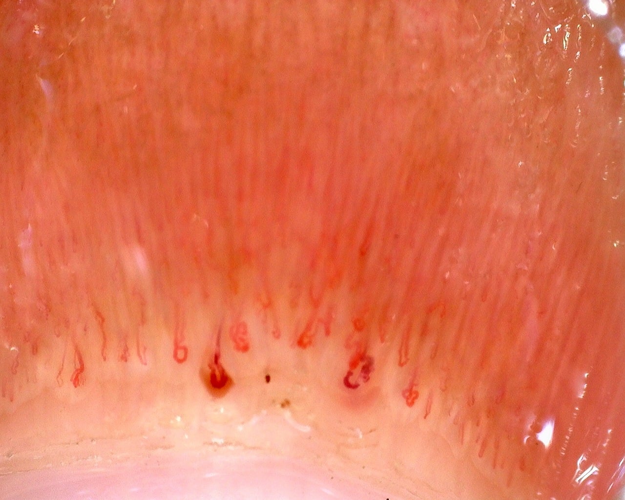
Nailfold overview from the juvenile dermatomyositis case with Gottron papules. The complete report describes the vascular abnormalities; this image is an orientation view.
Source study context
The baseline capillaroscopy is compatible with juvenile dermatomyositis and shows a severe scleroderma-like microangiopathy, with marked capillary loss, frequent enlarged and giant capillaries, hemorrhages, abnormal or arborizing shapes, and architectural disorganization.
Measurements and classification refer to the described study or visit; they are not new measurements of this image.
View the complete case and studyImage details
- Pathology
- Juvenile Dermatomyositis
- Pattern
- Late Scleroderma-Like Pattern
- Author or contributor
- Clara Udaondo
- Magnification
- 50×
- Device
- Dino-Lite Edge
- Field of view (W × H)
- ≈ 7.24 × 5.79 mm
Teaching point
Confirm that the first capillary row is visible and reasonably focused.
Clinical caveat
This image illustrates a visual finding and should be interpreted with acquisition quality, distribution across fingers, clinical context, and serology when relevant.
Use and licensing
Contributed by Clara Udaondo for educational publication on Capillaroscopy.com. For reuse permissions, contact the rights holder.
Related images
Back to atlasThere are not enough related images in this library yet.