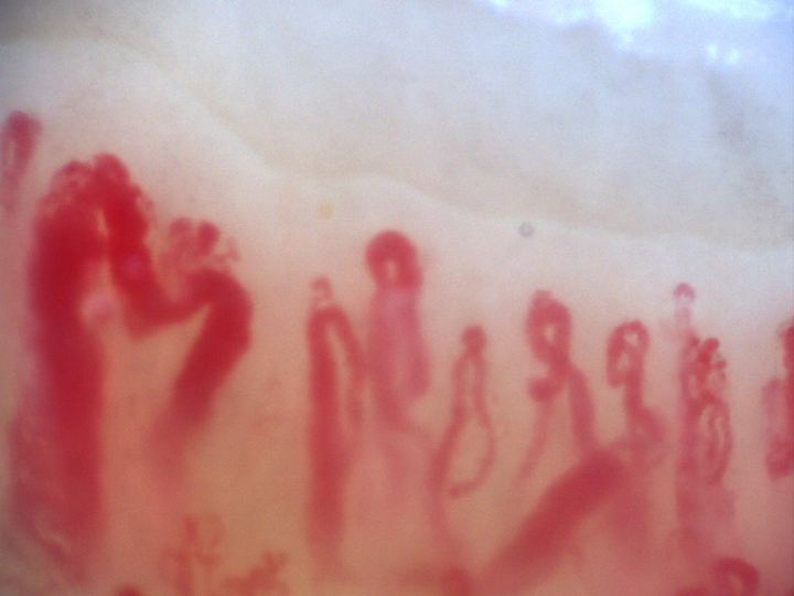
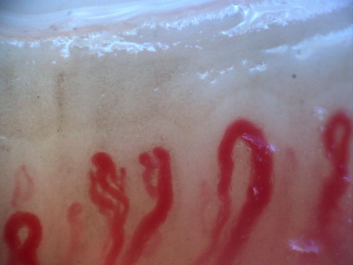
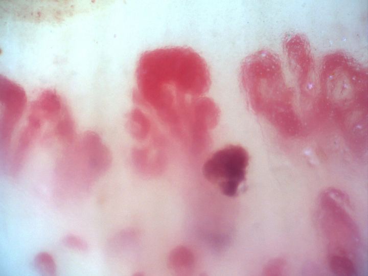
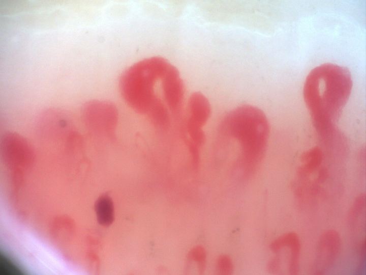
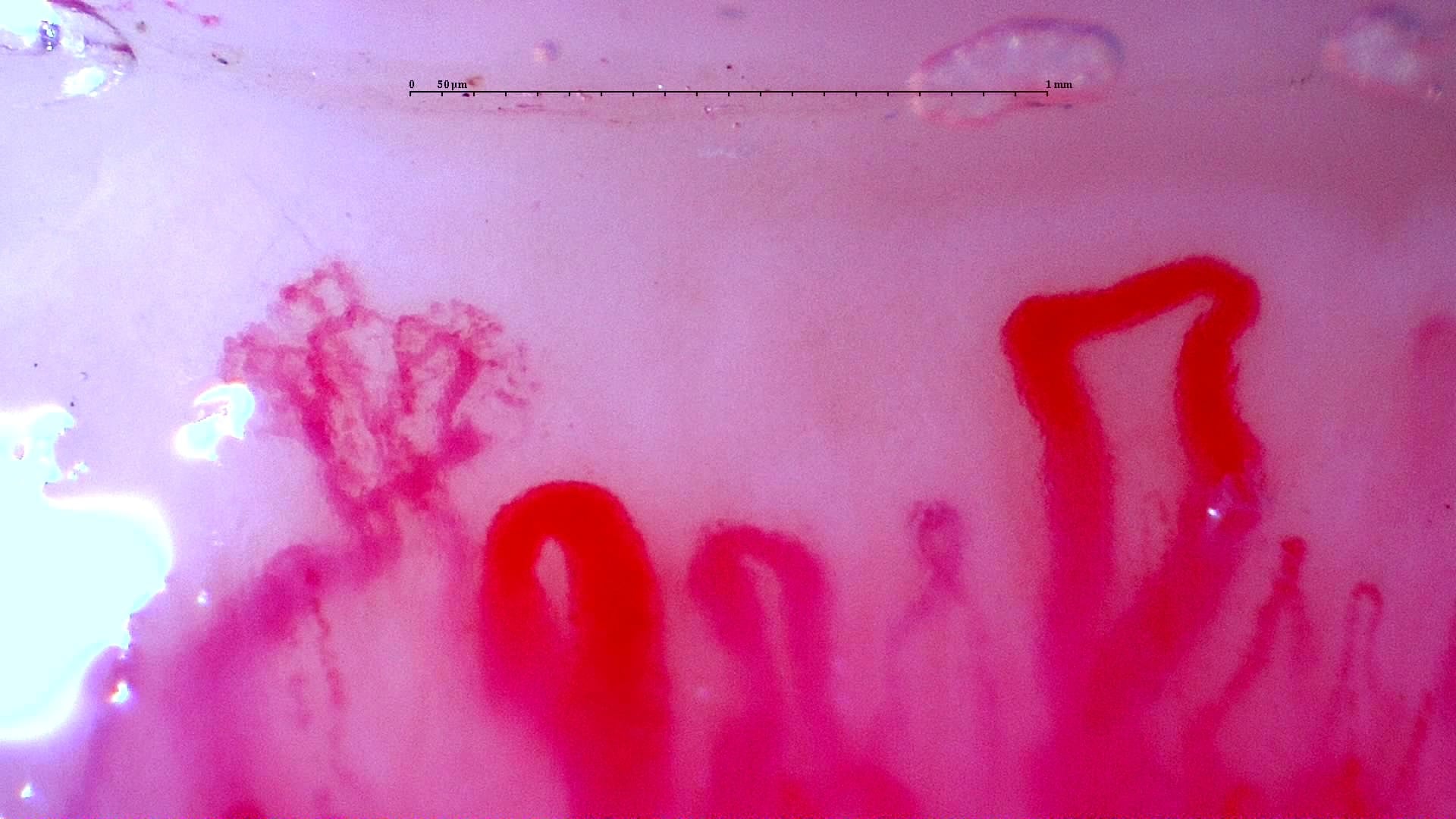
Anti-NXP2 dermatomyositis: initial enlarged loops
Prominent enlarged loops and a complex branching form in the initial anti-NXP2 dermatomyositis study. Compare this with the linked follow-up image and complete serial studies.
Source study context
The first study shows an active scleroderma/myositis-like microangiopathy with severe capillary loss, giant capillaries, arborizing ramifications, hemorrhages, architectural disorganization, and avascular areas. The follow-up study shows regression to a non-specific pattern with improved density and disappearance of giant capillaries and hemorrhages.
Measurements and classification refer to the described study or visit; they are not new measurements of this image.
View the complete case and studyImage details
- Pathology
- Dermatomyositis (anti-NXP2+)
- Pattern
- Myositis-Associated Microangiopathy with Treatment Response
- Author or contributor
- Luis Saez Comet
- Hallmarks
- Giant capillaryCapillary density
- Magnification
- 250×
- Device
- Smart G-Scope
- Field of view (W × H)
- ≈ 2.30 × 1.29 mm
Teaching point
Describe whether abnormalities are isolated, repeated, or combined.
Clinical caveat
This image illustrates a visual finding and should be interpreted with acquisition quality, distribution across fingers, clinical context, and serology when relevant.
Use and licensing
Contributed by Luis Saez Comet for educational publication on Capillaroscopy.com. For reuse permissions, contact the rights holder.