Interactive viewer
Anti-TIF1γ Dermatomyositis with Scleroderma-Like Pattern
Images & measurements22The study, field by field
Images & measurements
Explore the fields from each finger and open any image for a closer look. Measurements describe the complete examination at each visit.
Contributed by Franklin Uguña Sari for educational publication on Capillaroscopy.com. For reuse permissions, contact the rights holder.
Anti-TIF1γ Dermatomyositis with Scleroderma-Like Pattern
Capillaroscopy shows severely reduced capillary density (2.7/mm), architectural disorganization, frequent dilations, giant capillaries, and abnormal/ramified forms (2/3 on the semi-quantitative scale), consistent with a scleroderma-like pattern.
Recorded measurements
- Capillary density
- 2.73capillaries/mm
- Enlarged capillaries
- 41.11%
- Giant capillaries
- 36.67%
- Hemorrhages
- 0.03/mm
Values from the saved analysis: density and hemorrhages per total analyzed length; percentages of the total capillary count. These are not measurements of an individual image and may differ from averages across fields.
Left index finger
6 images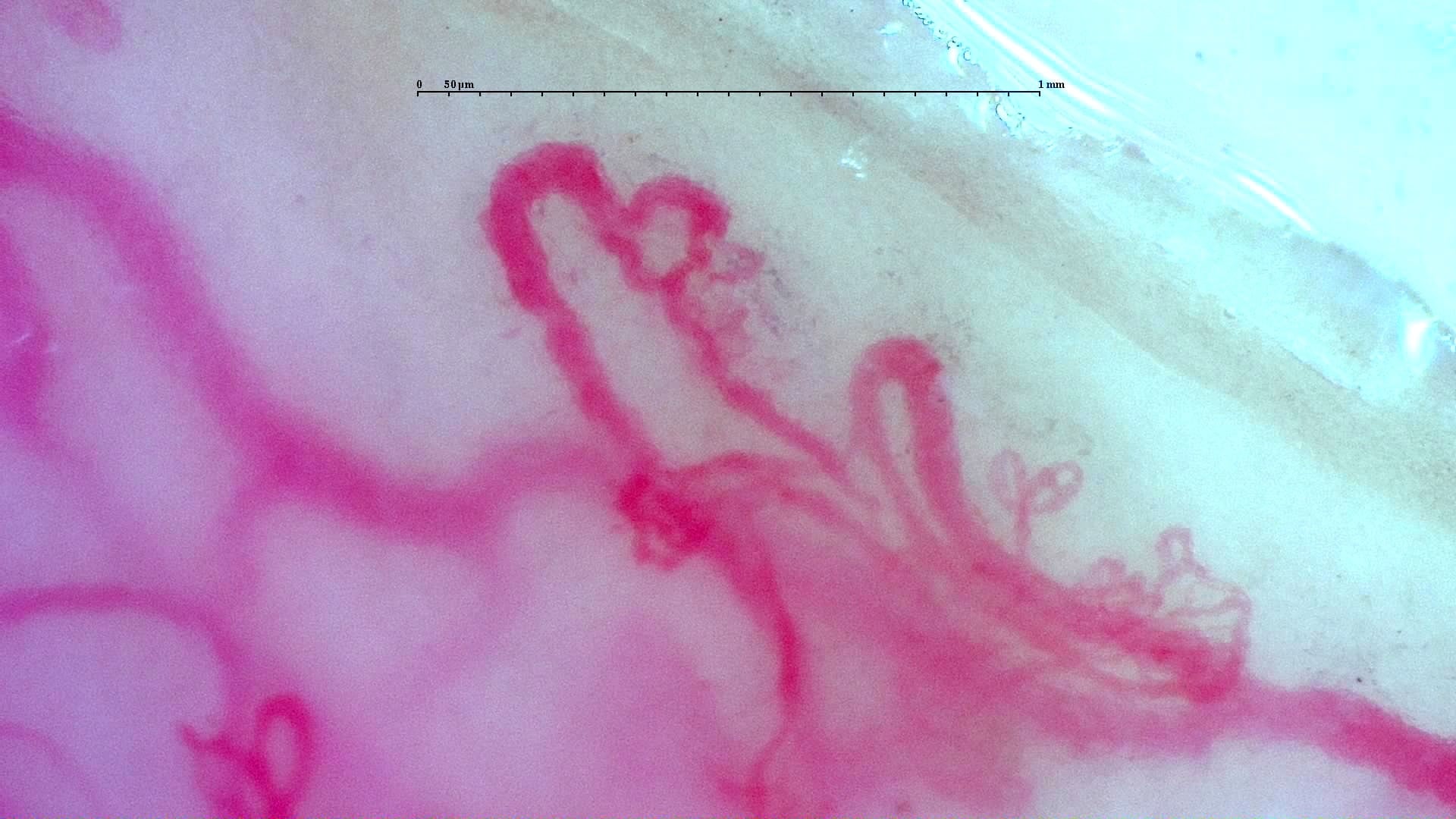
Nailfold capillaroscopy: left index finger, field b.
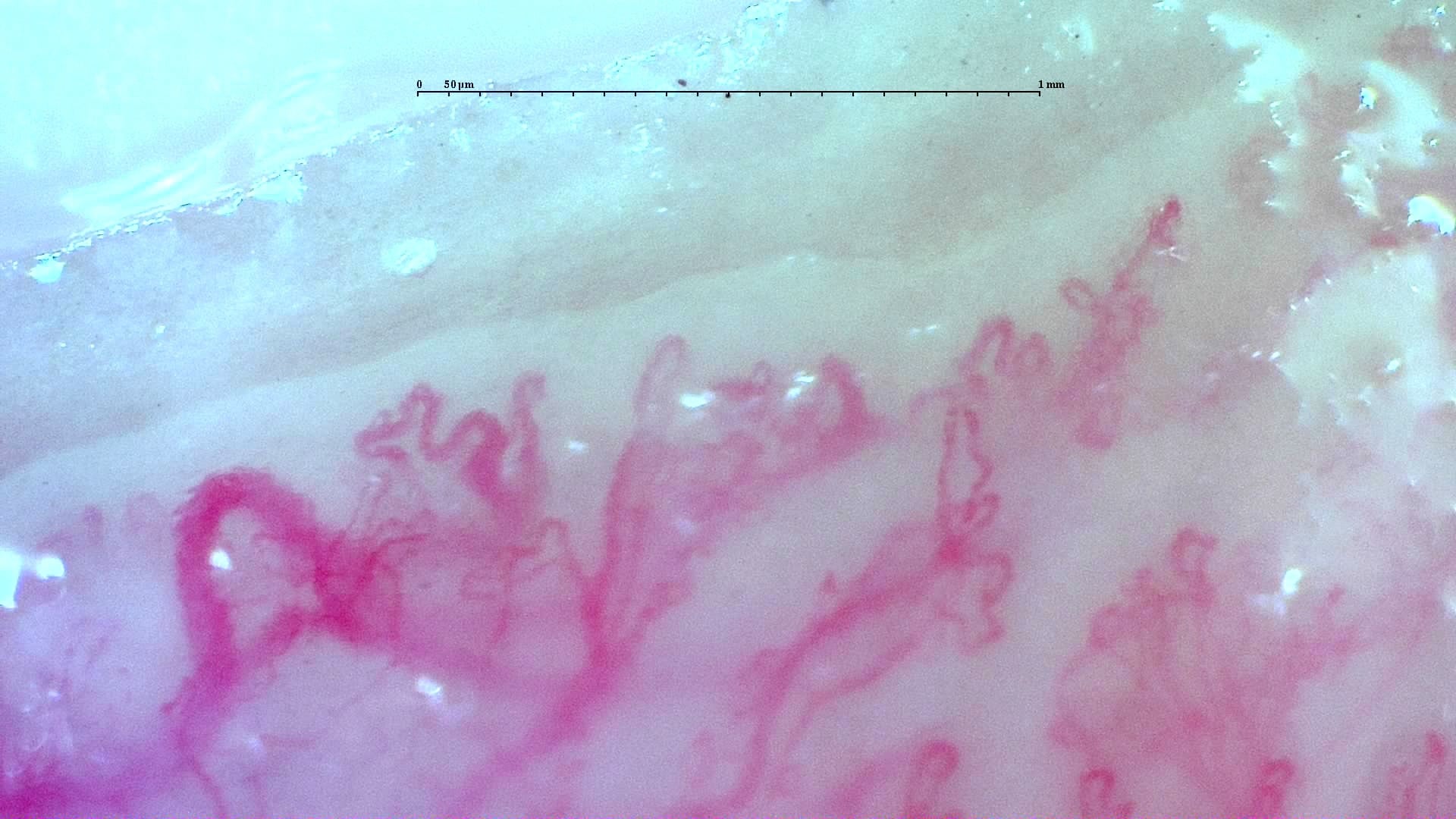
Nailfold capillaroscopy: left index finger, field b.
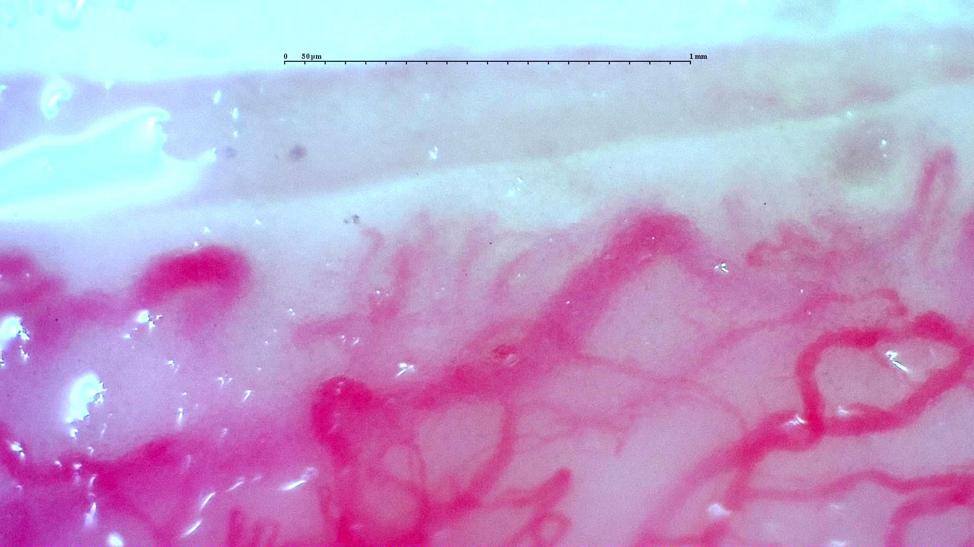
Nailfold capillaroscopy: left index finger, field c.
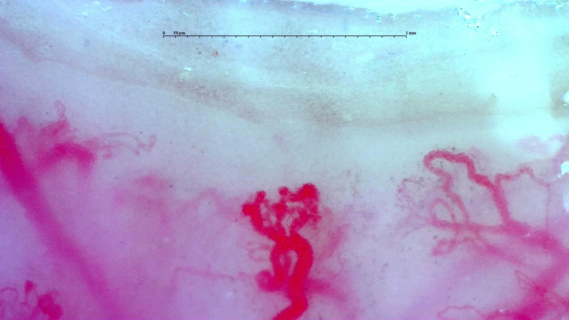
Nailfold capillaroscopy: left index finger, field c.
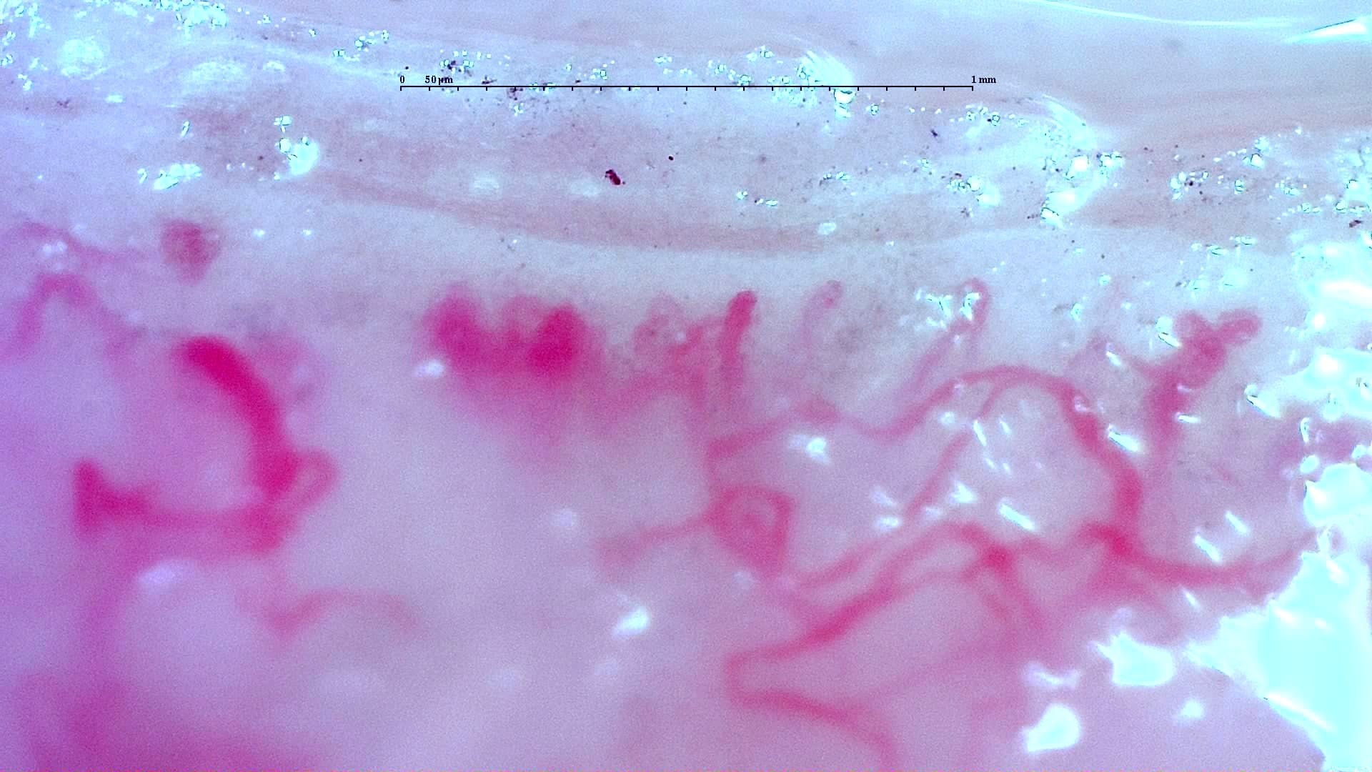
Nailfold capillaroscopy: left index finger, field c.
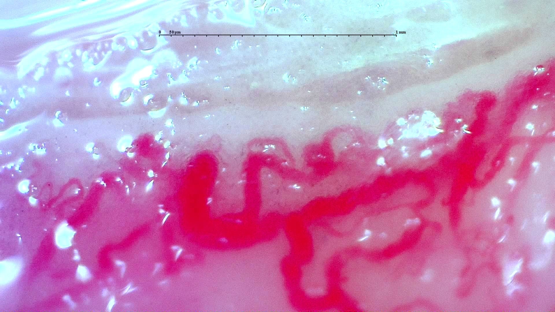
Nailfold capillaroscopy: left index finger, field d.
Left middle finger
3 images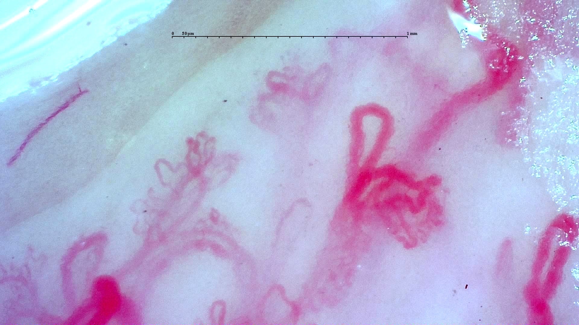
Nailfold capillaroscopy: left middle finger, field a.
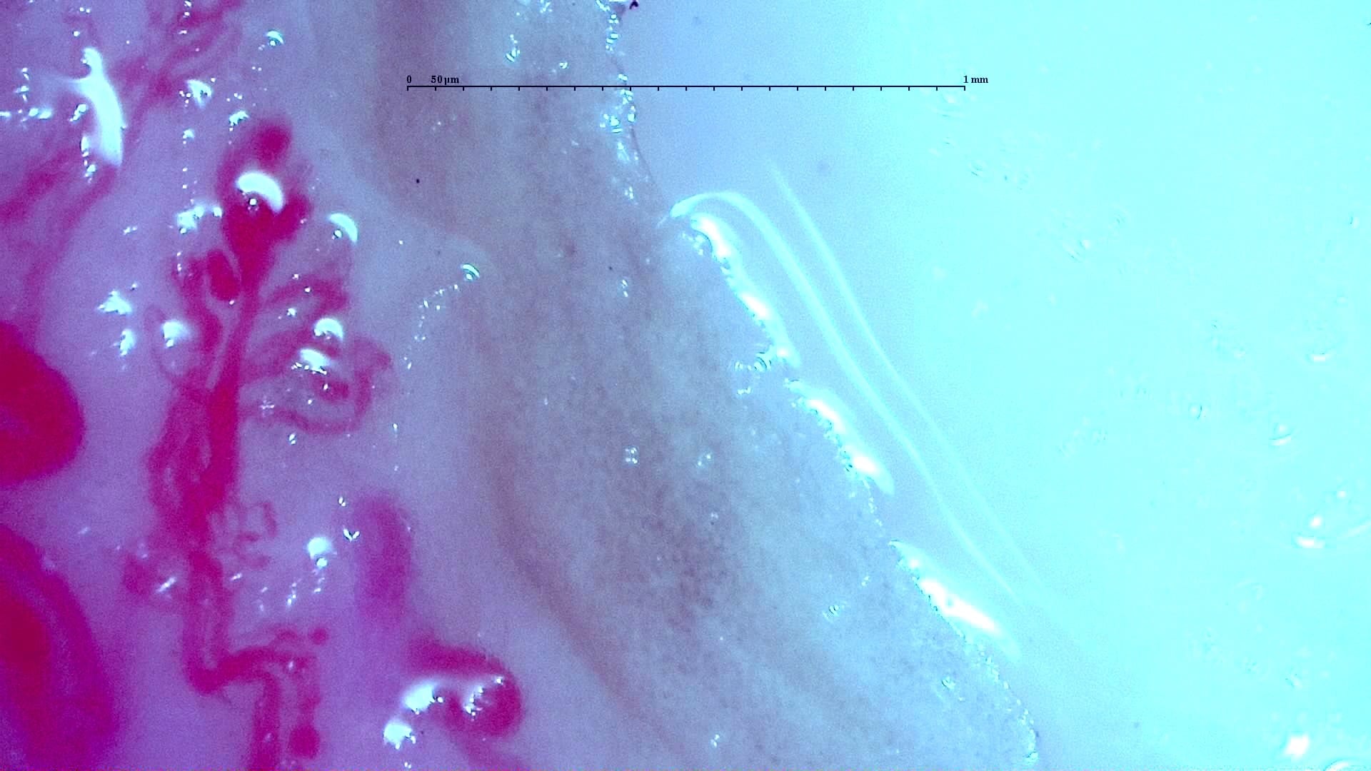
Nailfold capillaroscopy: left middle finger, field b.
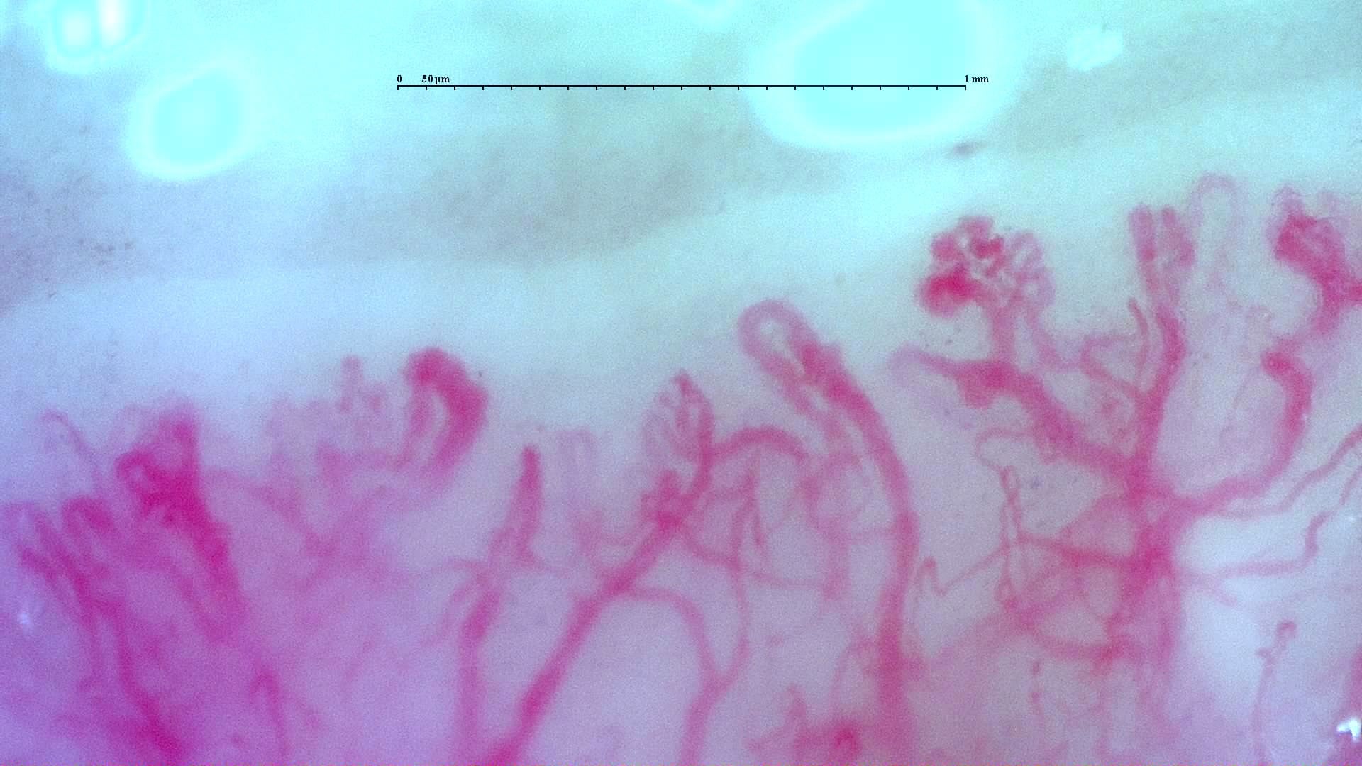
Complex, winding and ramified vascular forms in the anti-TIF1γ dermatomyositis case. A scleroderma-like image appearance does not establish systemic sclerosis.
Read the atlas commentaryLeft ring finger
1 images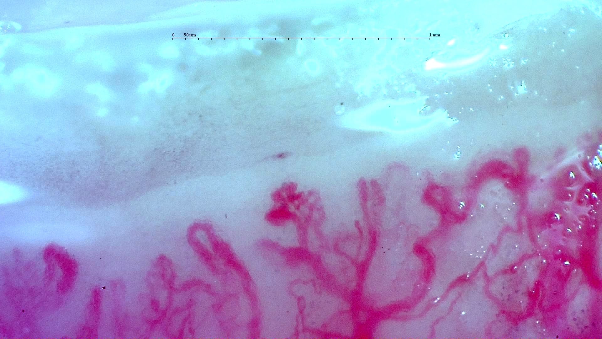
Nailfold capillaroscopy: left ring finger, field b.
Left little finger
1 images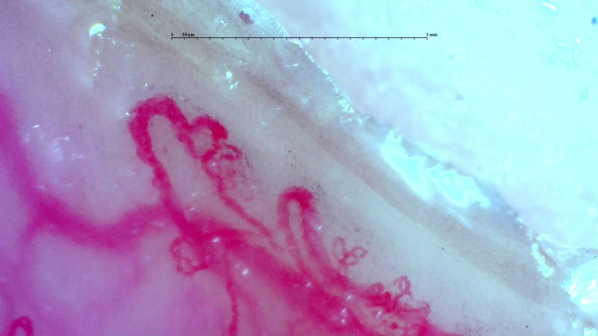
Nailfold capillaroscopy: left little finger, field d.
Right index finger
4 images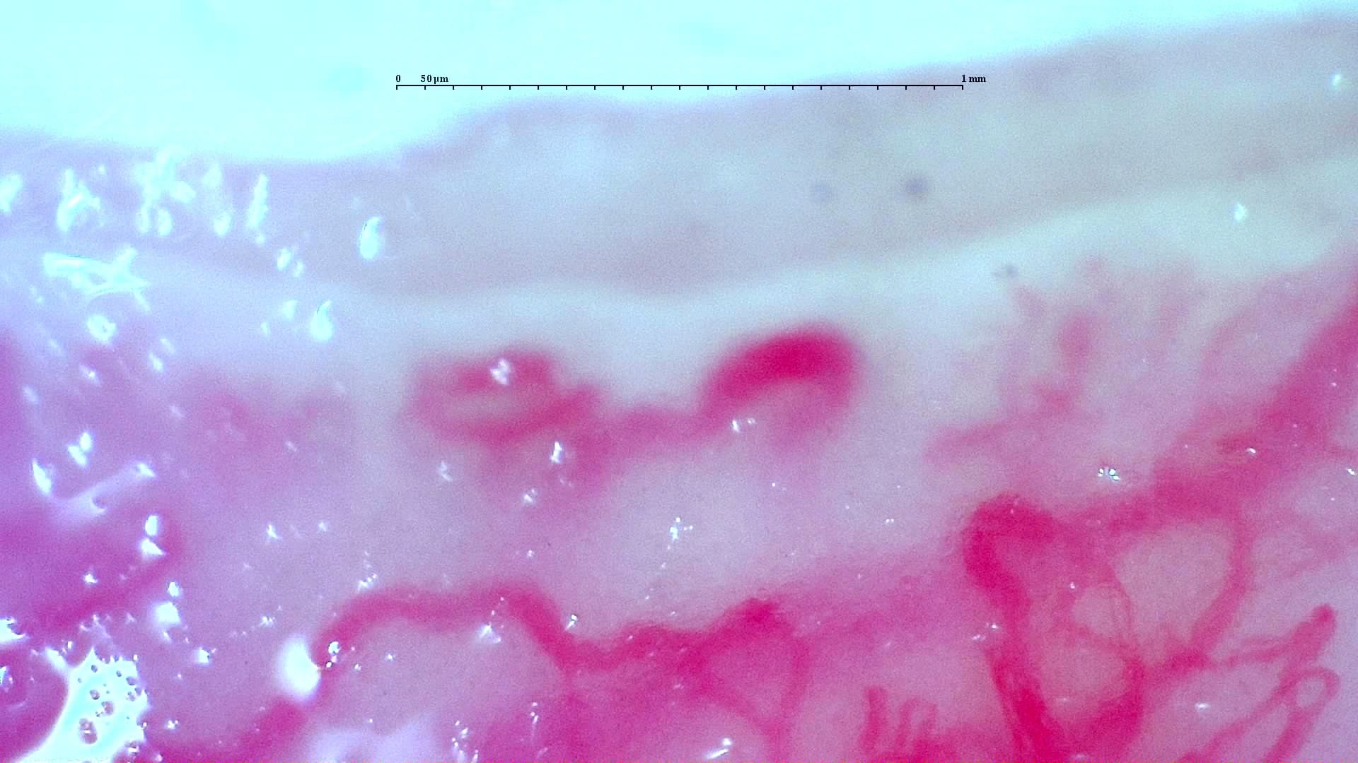
Nailfold capillaroscopy: right index finger, field b.
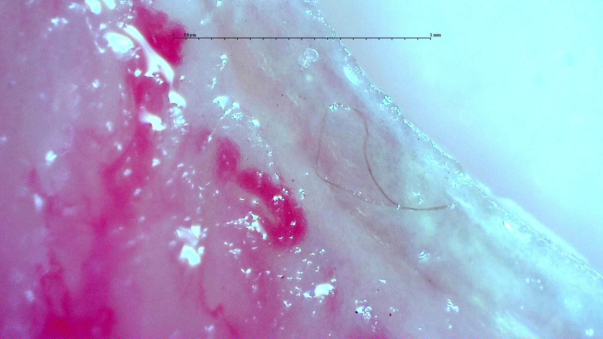
Nailfold capillaroscopy: right index finger, field b.
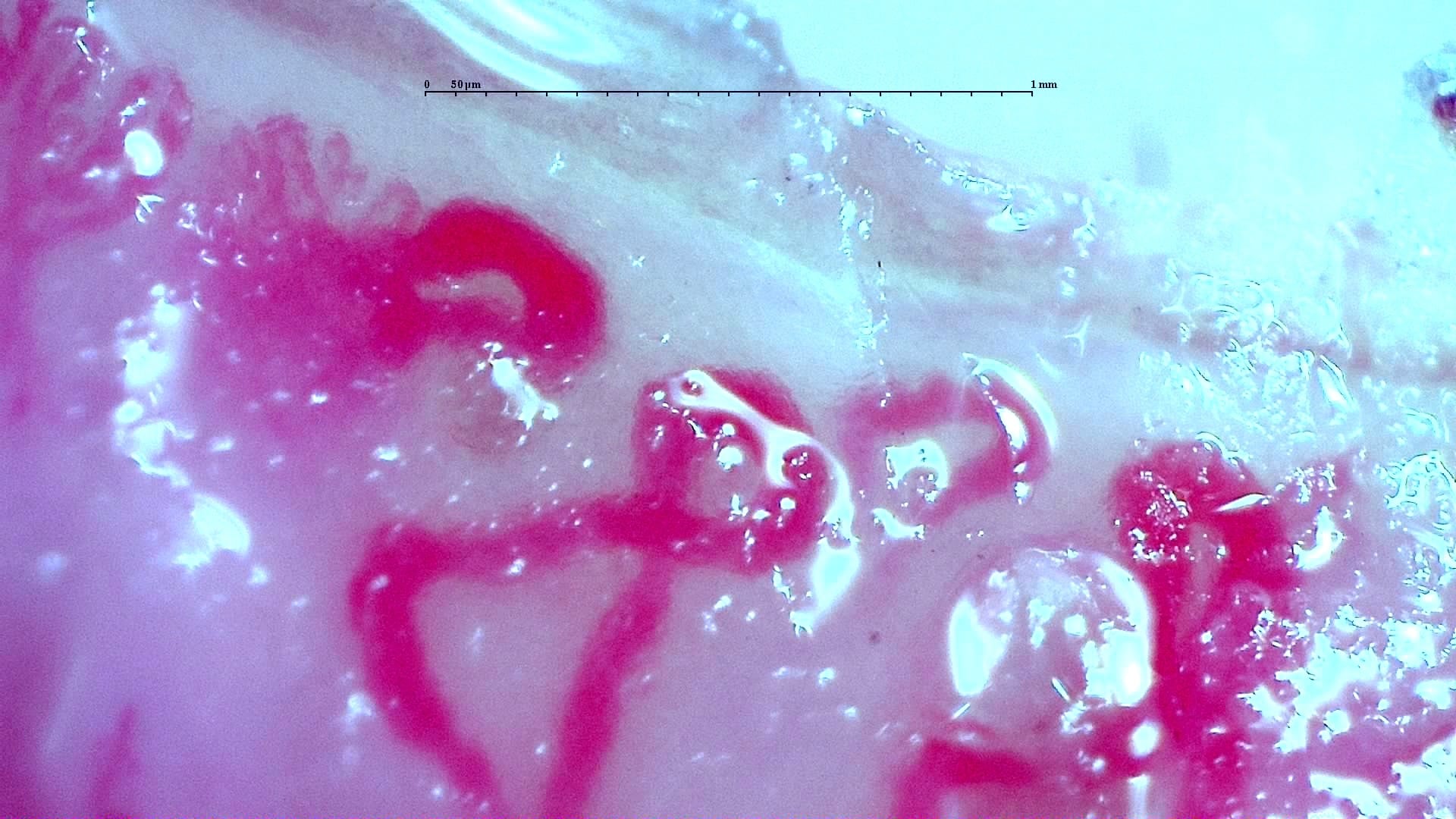
Nailfold capillaroscopy: right index finger, field c.
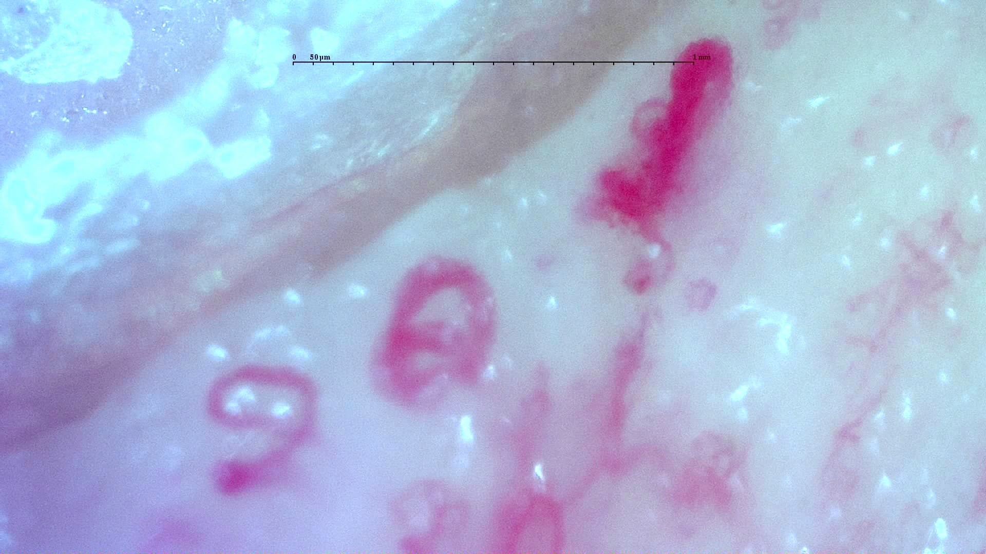
Nailfold capillaroscopy: right index finger, field c.
Right middle finger
4 images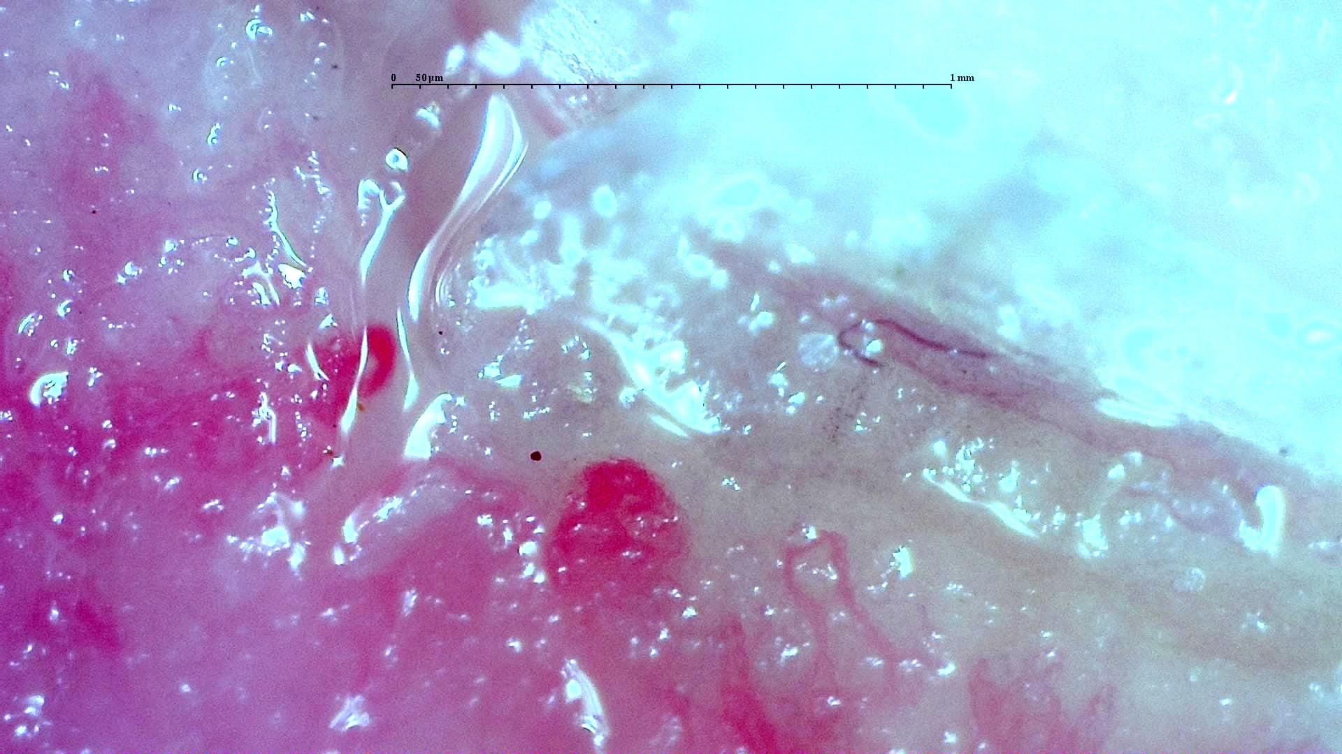
Nailfold capillaroscopy: right middle finger, field b.
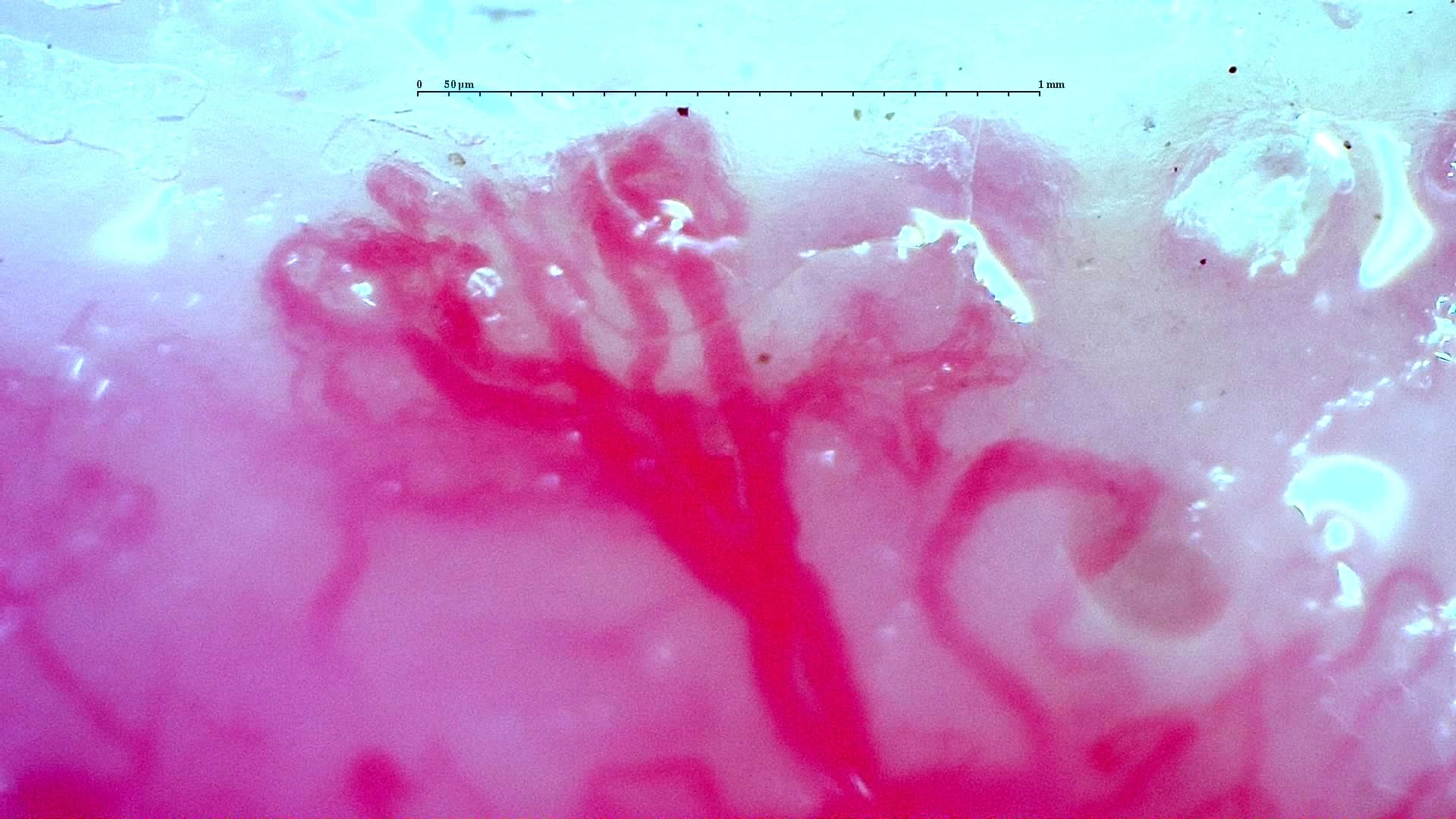
Nailfold capillaroscopy: right middle finger, field c.
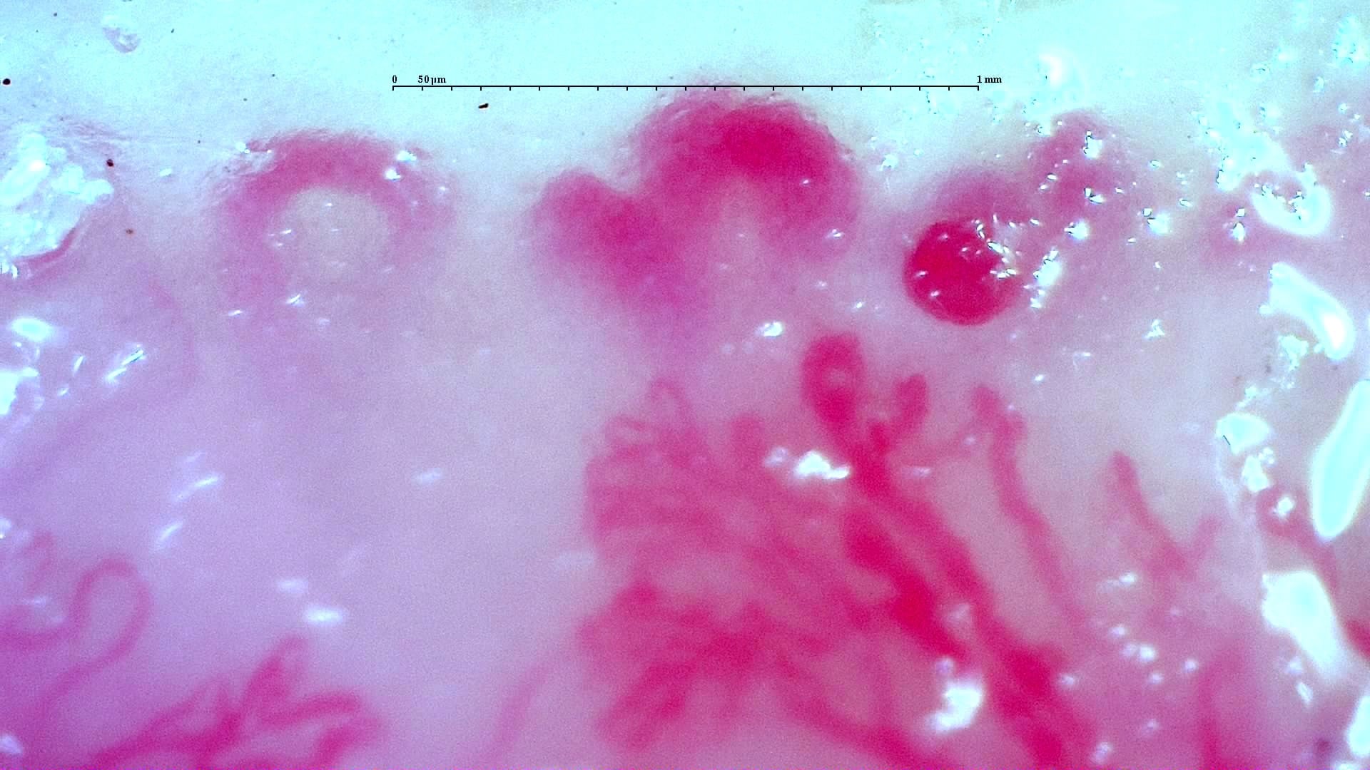
Nailfold capillaroscopy: right middle finger, field c.
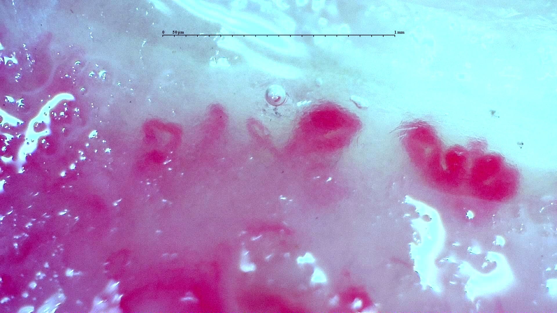
Nailfold capillaroscopy: right middle finger, field d.
Right ring finger
1 images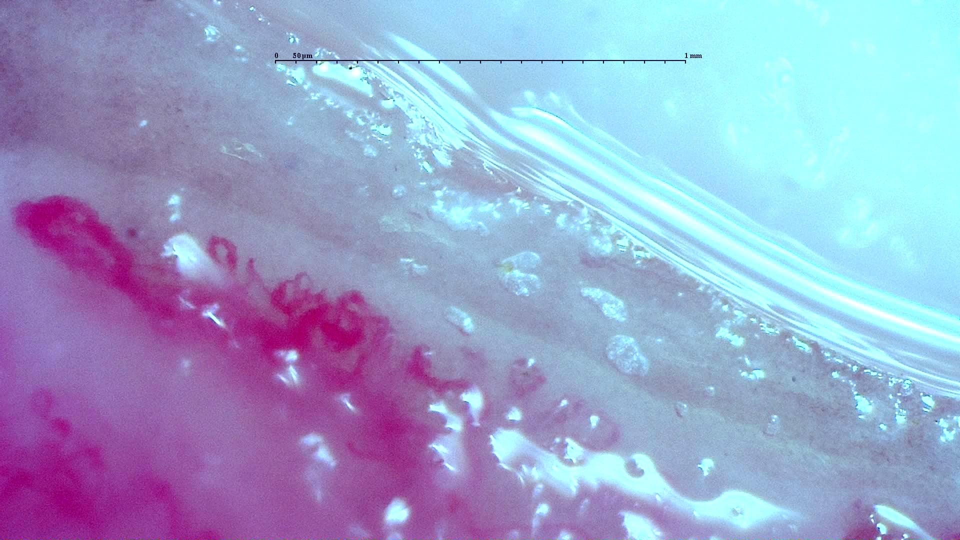
Nailfold capillaroscopy: right ring finger, field c.
Right little finger
2 images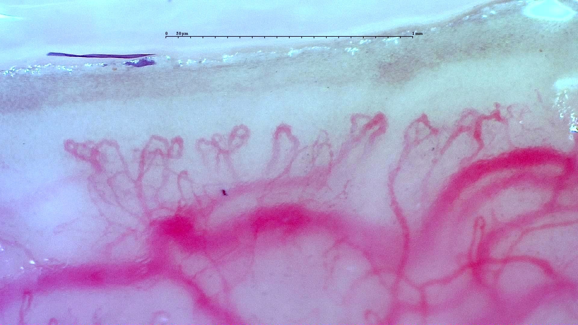
Nailfold capillaroscopy: right little finger, field a.
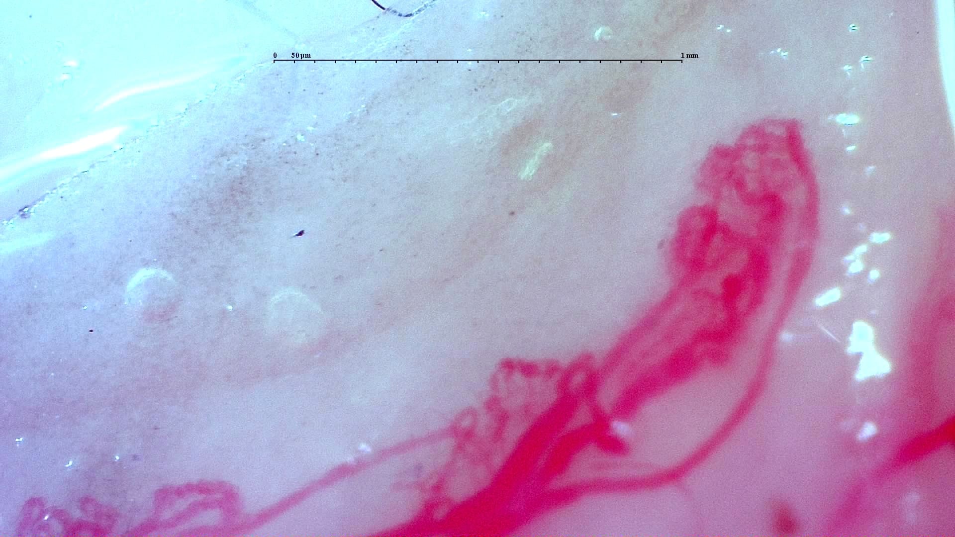
Nailfold capillaroscopy: right little finger, field b.