Interactive viewer
Mixed Connective Tissue Disease with an Unclassifiable Scleroderma-Like Pattern
Images & measurements25The study, field by field
Images & measurements
Explore the fields from each finger and open any image for a closer look. Measurements describe the complete examination at each visit.
Contributed by Miguel Antonio Mesa Navas for educational publication on Capillaroscopy.com. For reuse permissions, contact the rights holder.
Mixed Connective Tissue Disease with an Unclassifiable Scleroderma-Like Pattern
Capillaroscopy shows megacapillaries, but their distribution does not fit the typical organization of any early, active, or late Cutolo subtype. The overall morphology is therefore classified as a scleroderma-like pattern. Arborizing capillaries are also evident. The quantitative dataset reports a mean capillary density of 5.69/mm, enlarged capillaries in 78.7%, megacapillaries in 6.0%, and ramified forms in 4.7%.
Recorded measurements
- Capillary density
- 5.73capillaries/mm
- Enlarged capillaries
- 80.16%
- Giant capillaries
- 5.45%
- Hemorrhages
- 0.02/mm
Values from the saved analysis: density and hemorrhages per total analyzed length; percentages of the total capillary count. These are not measurements of an individual image and may differ from averages across fields.
Left index finger
2 images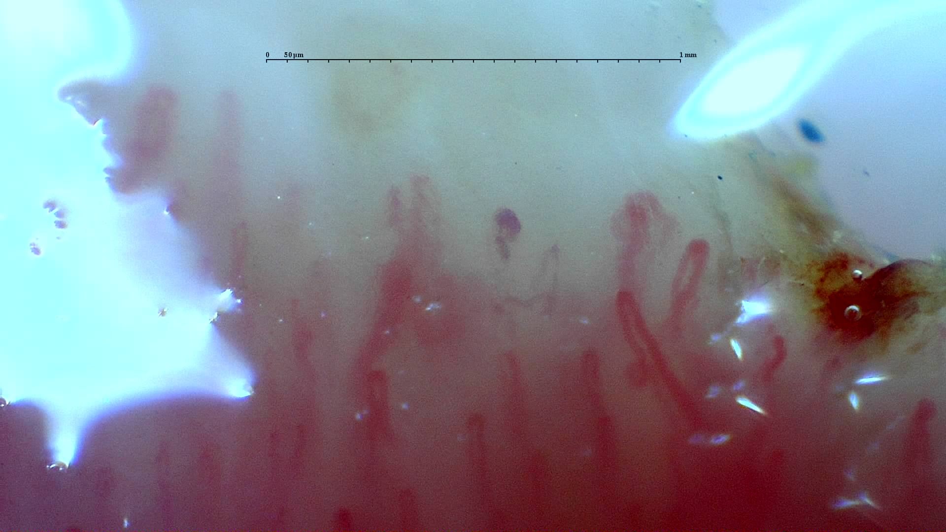
Nailfold capillaroscopy: left index finger, field d.
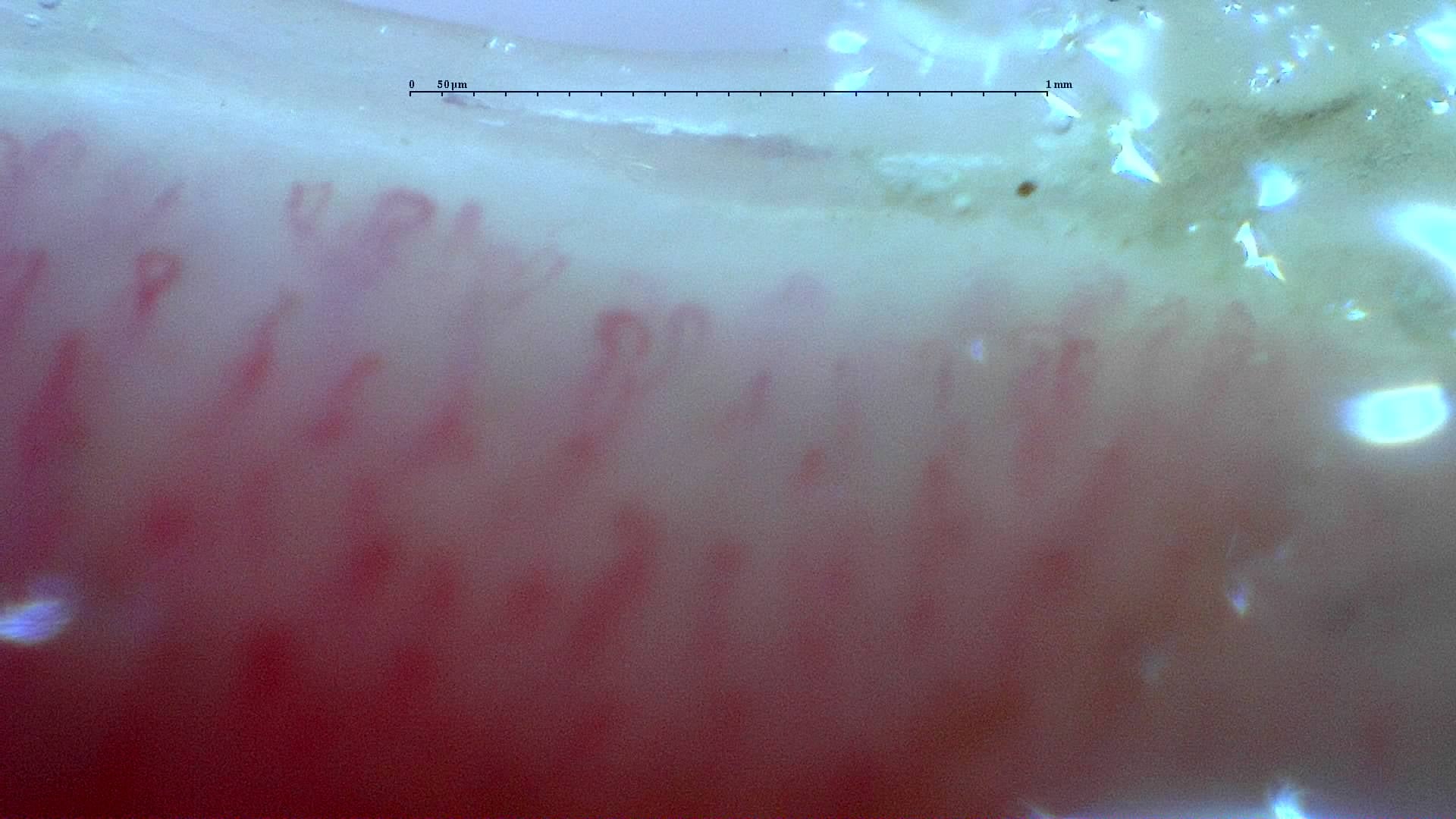
Nailfold capillaroscopy: left index finger, field b.
Left middle finger
4 images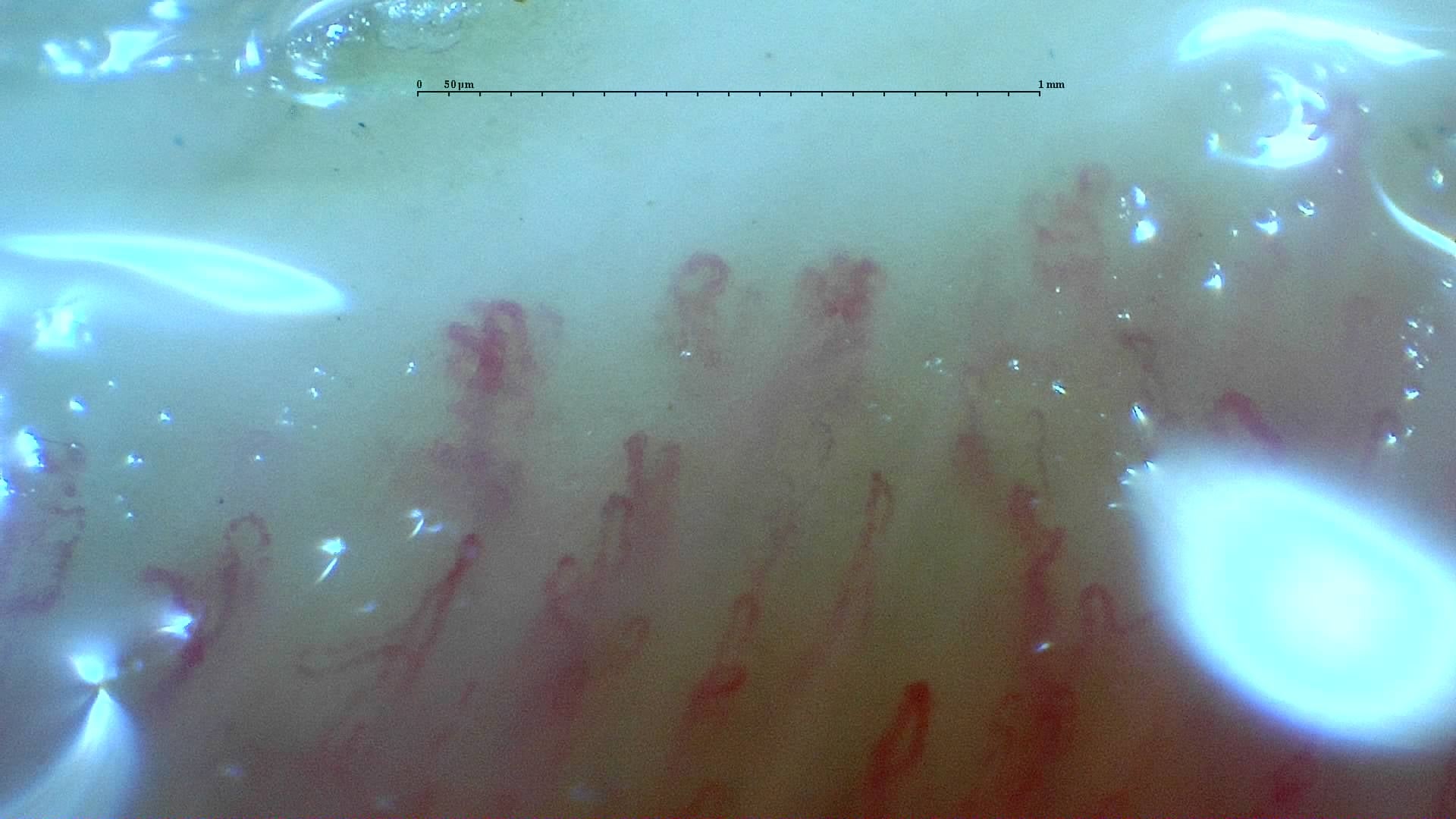
Nailfold capillaroscopy: left middle finger, field a.
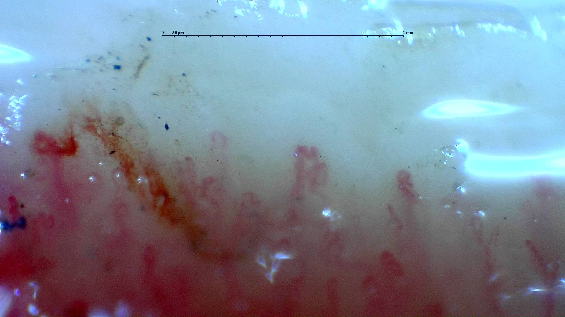
Nailfold capillaroscopy: left middle finger, field d.
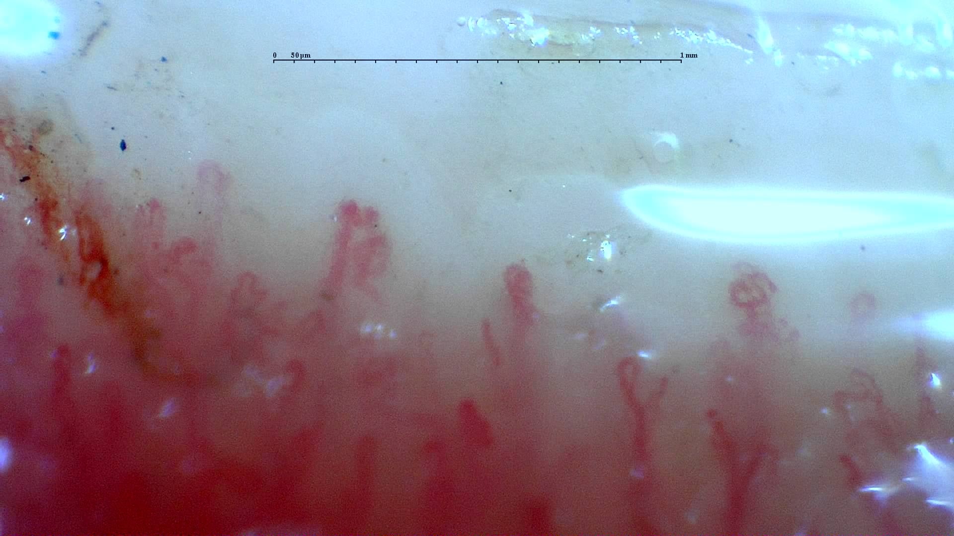
Nailfold capillaroscopy: left middle finger, field c.
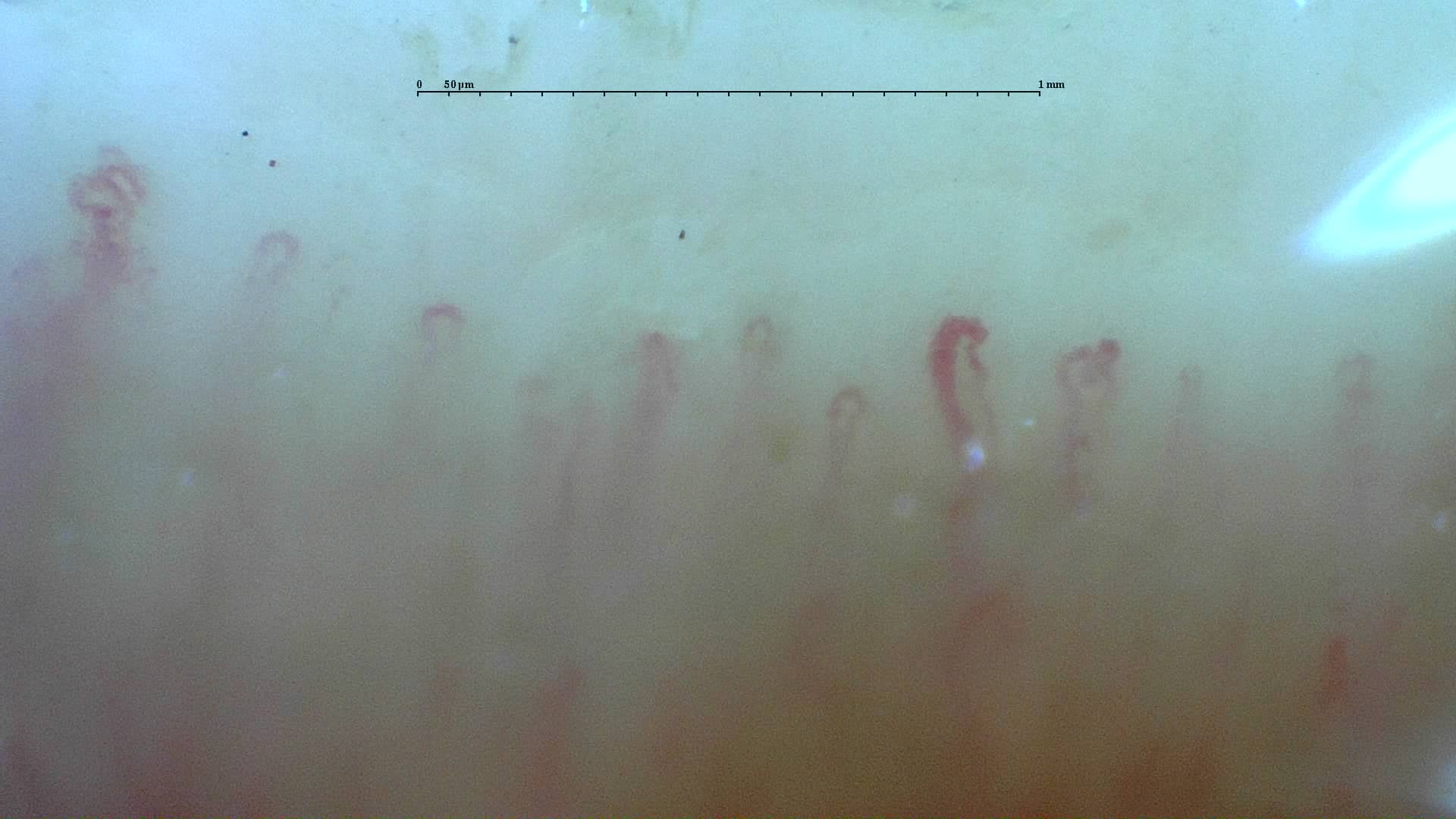
Nailfold capillaroscopy: left middle finger, field b.
Left ring finger
4 images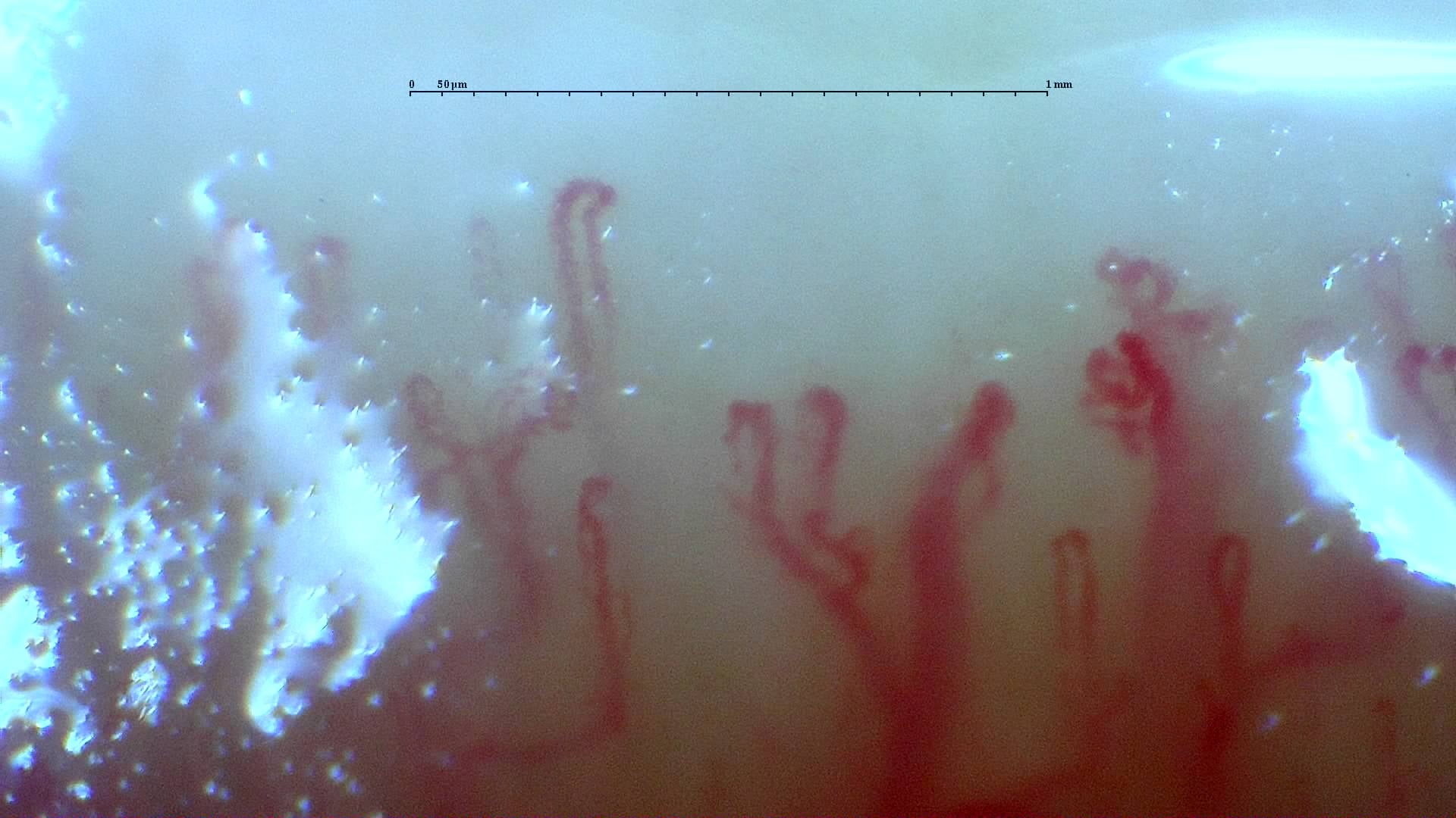
Nailfold capillaroscopy: left ring finger, field d.
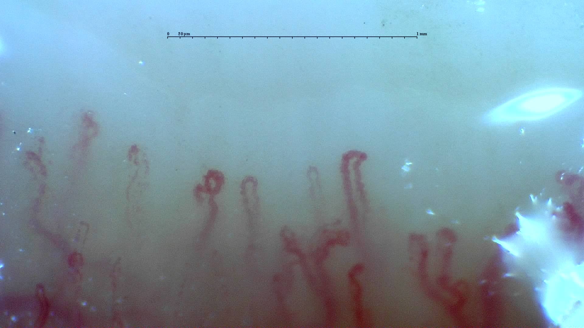
Nailfold capillaroscopy: left ring finger, field c.
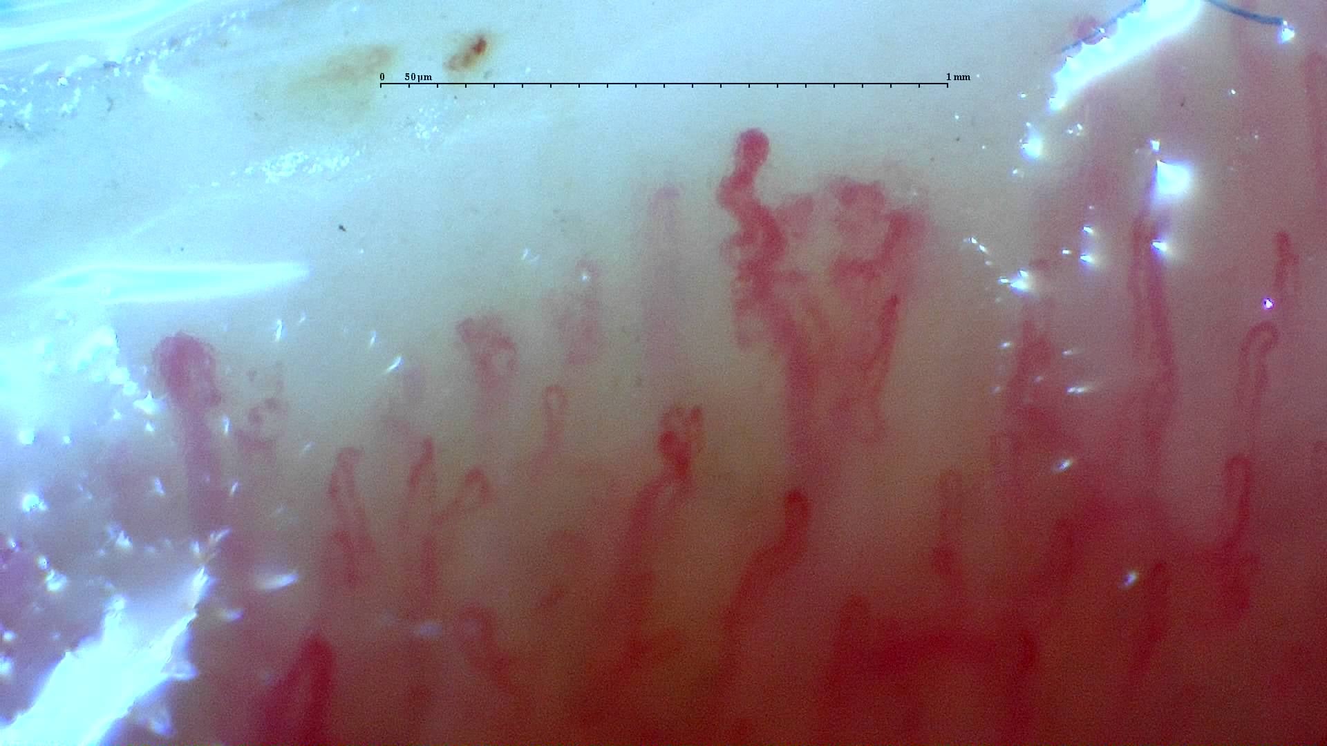
Nailfold capillaroscopy: left ring finger, field a.
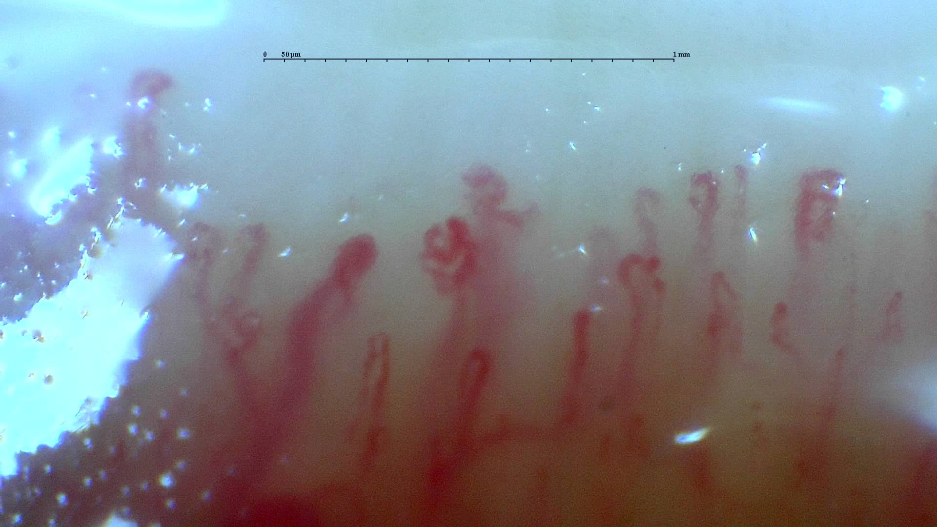
Irregular and branching forms in the anti-RNP-positive mixed connective tissue disease case. The source report describes a scleroderma-like pattern without an assignable Cutolo subtype.
Read the atlas commentaryLeft little finger
3 images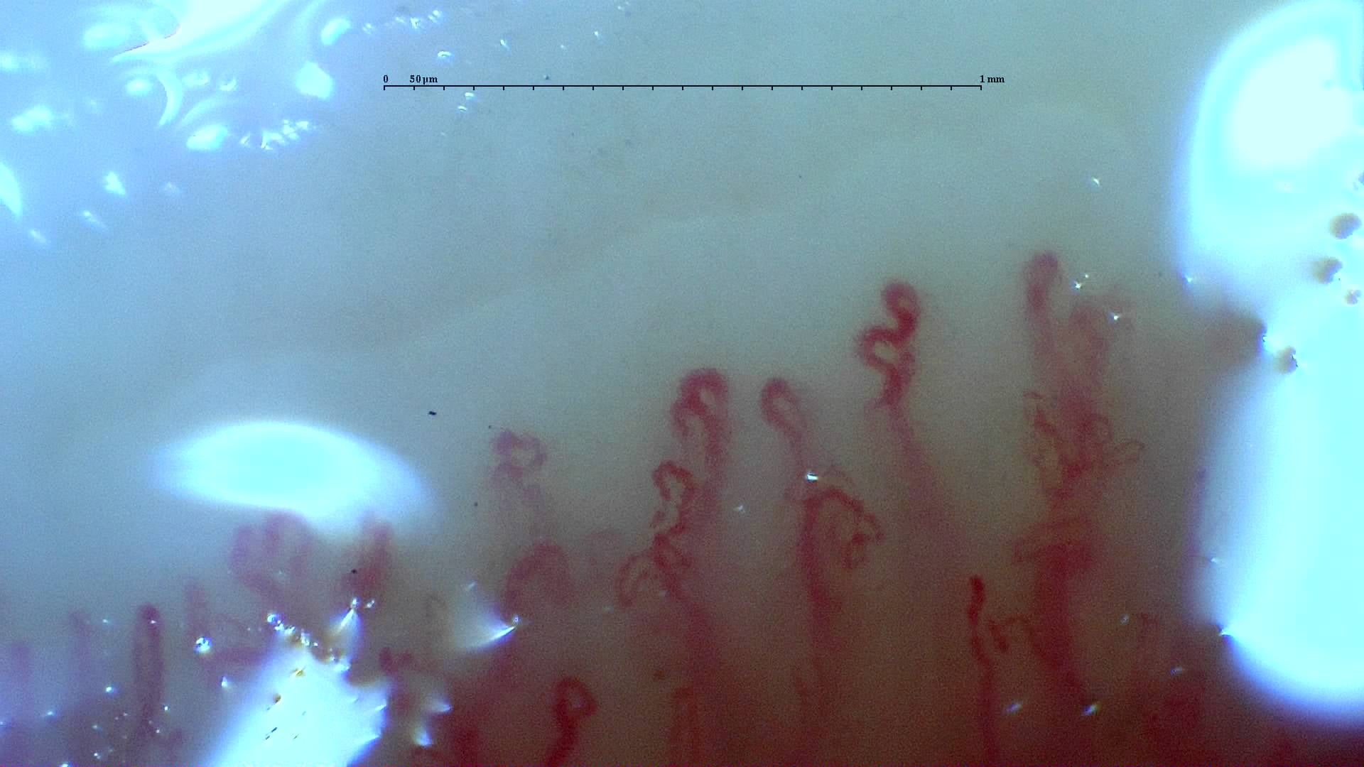
Nailfold capillaroscopy: left little finger, field a.
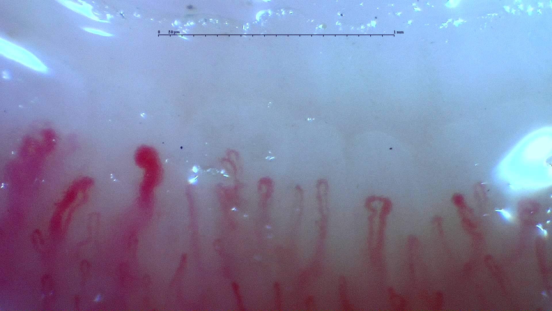
Nailfold capillaroscopy: left little finger, field d.
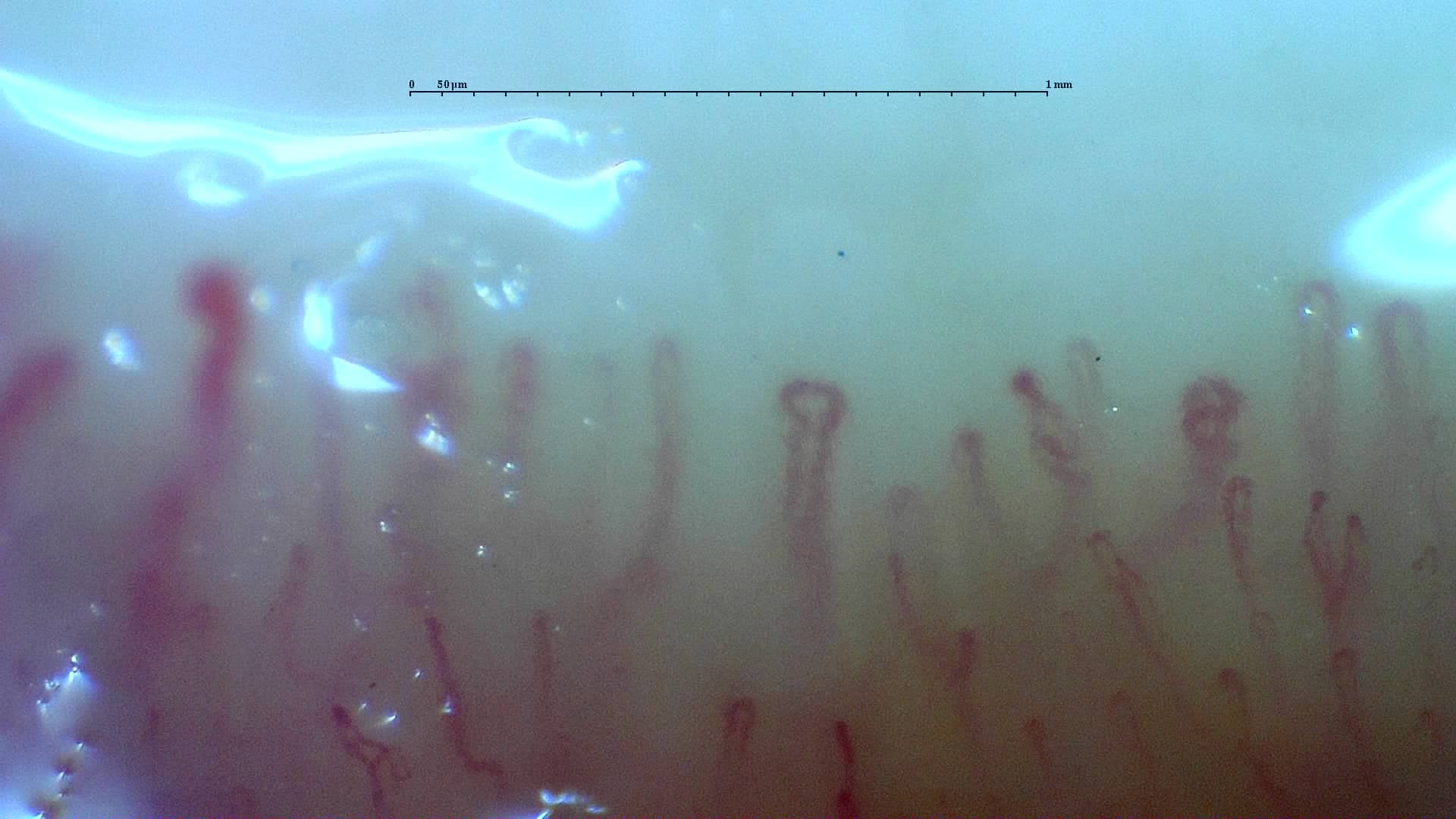
Nailfold capillaroscopy: left little finger, field c.
Right index finger
1 images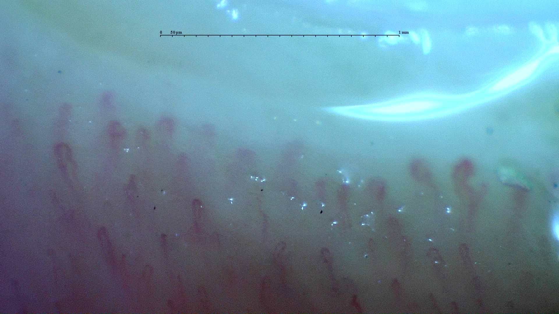
Nailfold capillaroscopy: right index finger, field d.
Right middle finger
4 images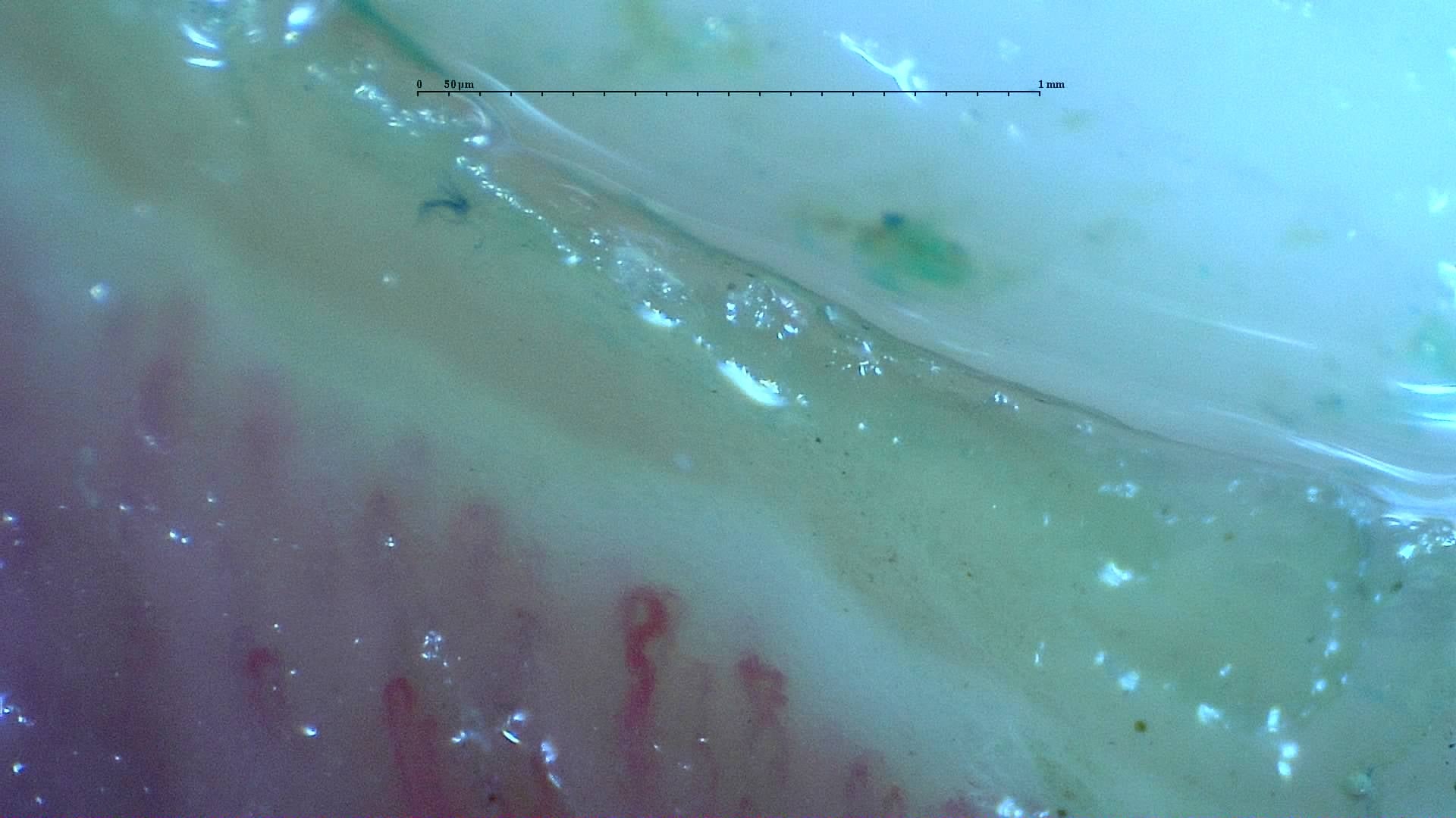
Nailfold capillaroscopy: right middle finger, field d.
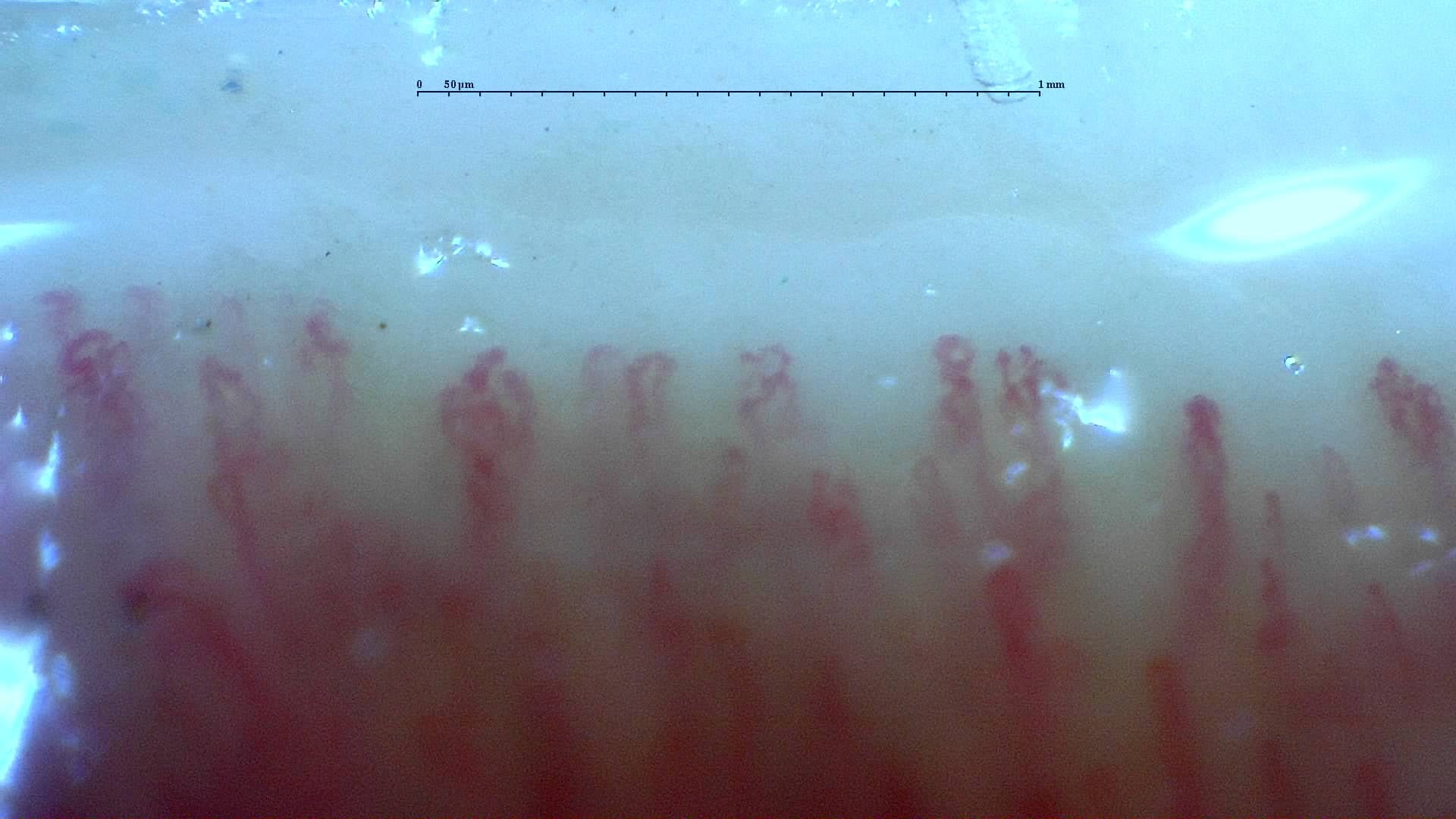
Nailfold capillaroscopy: right middle finger, field c.
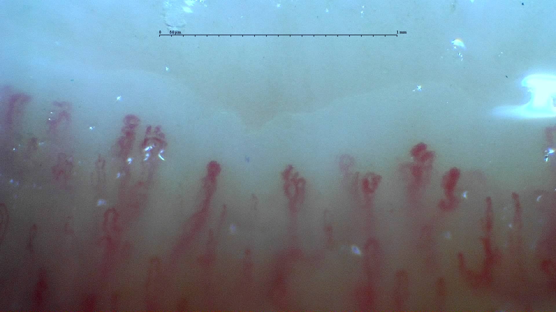
Nailfold capillaroscopy: right middle finger, field b.
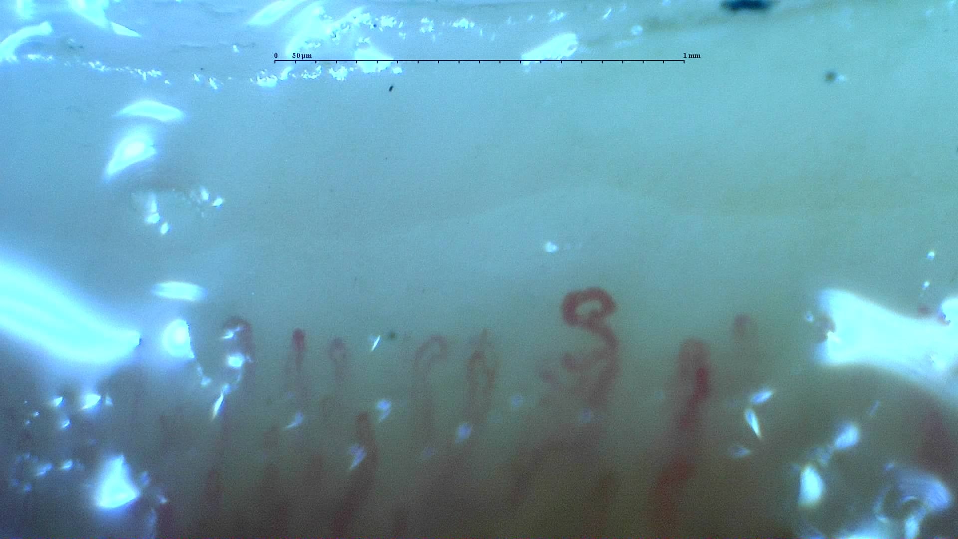
Nailfold capillaroscopy: right middle finger, field a.
Right ring finger
3 images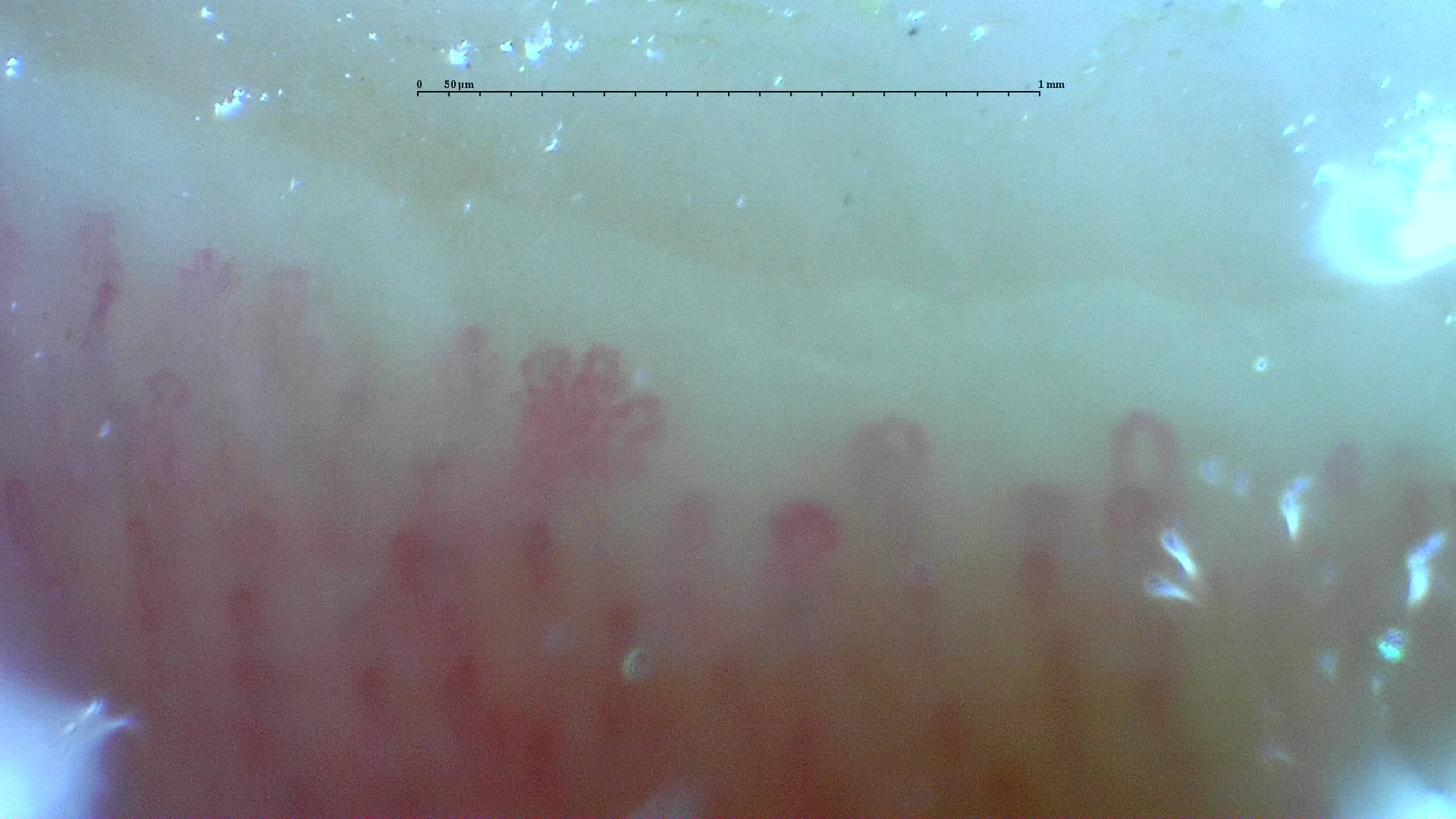
Nailfold capillaroscopy: right ring finger, field d.
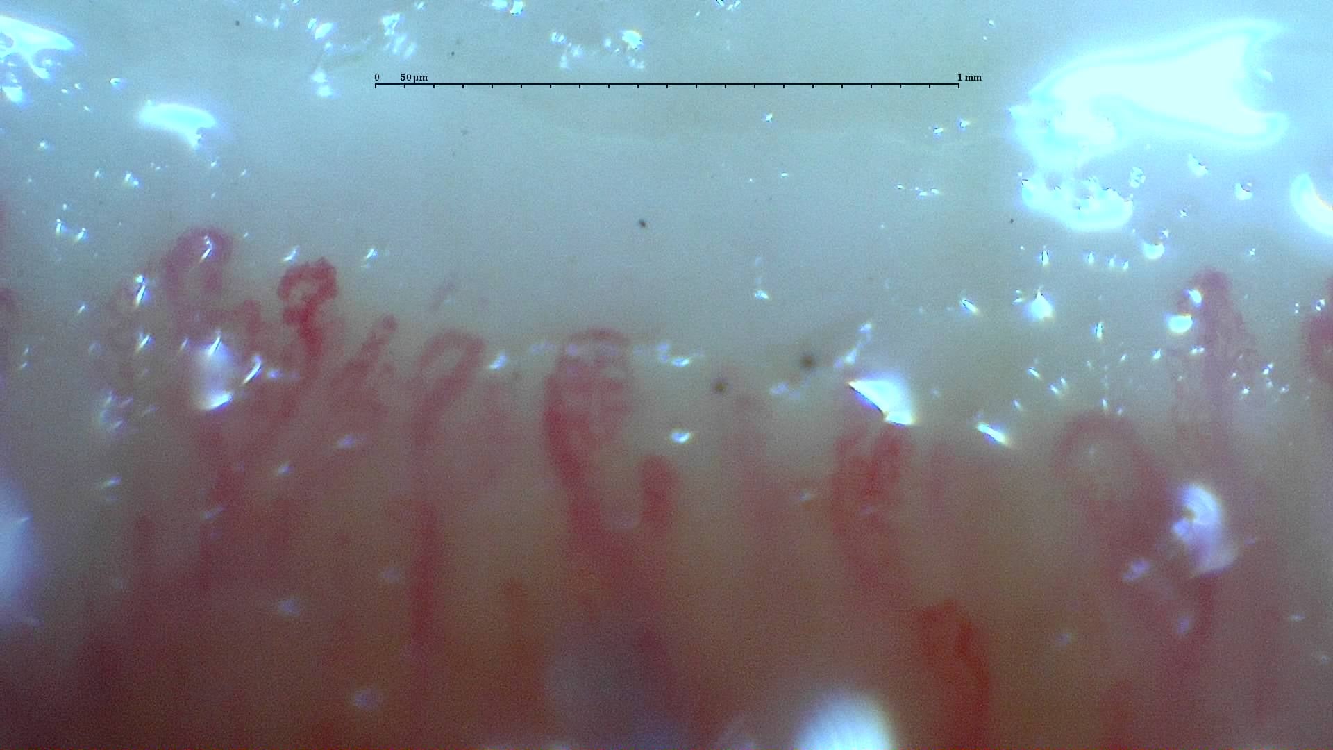
Nailfold capillaroscopy: right ring finger, field b.
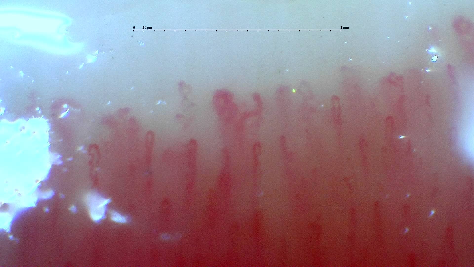
Nailfold capillaroscopy: right ring finger, field a.
Right little finger
4 images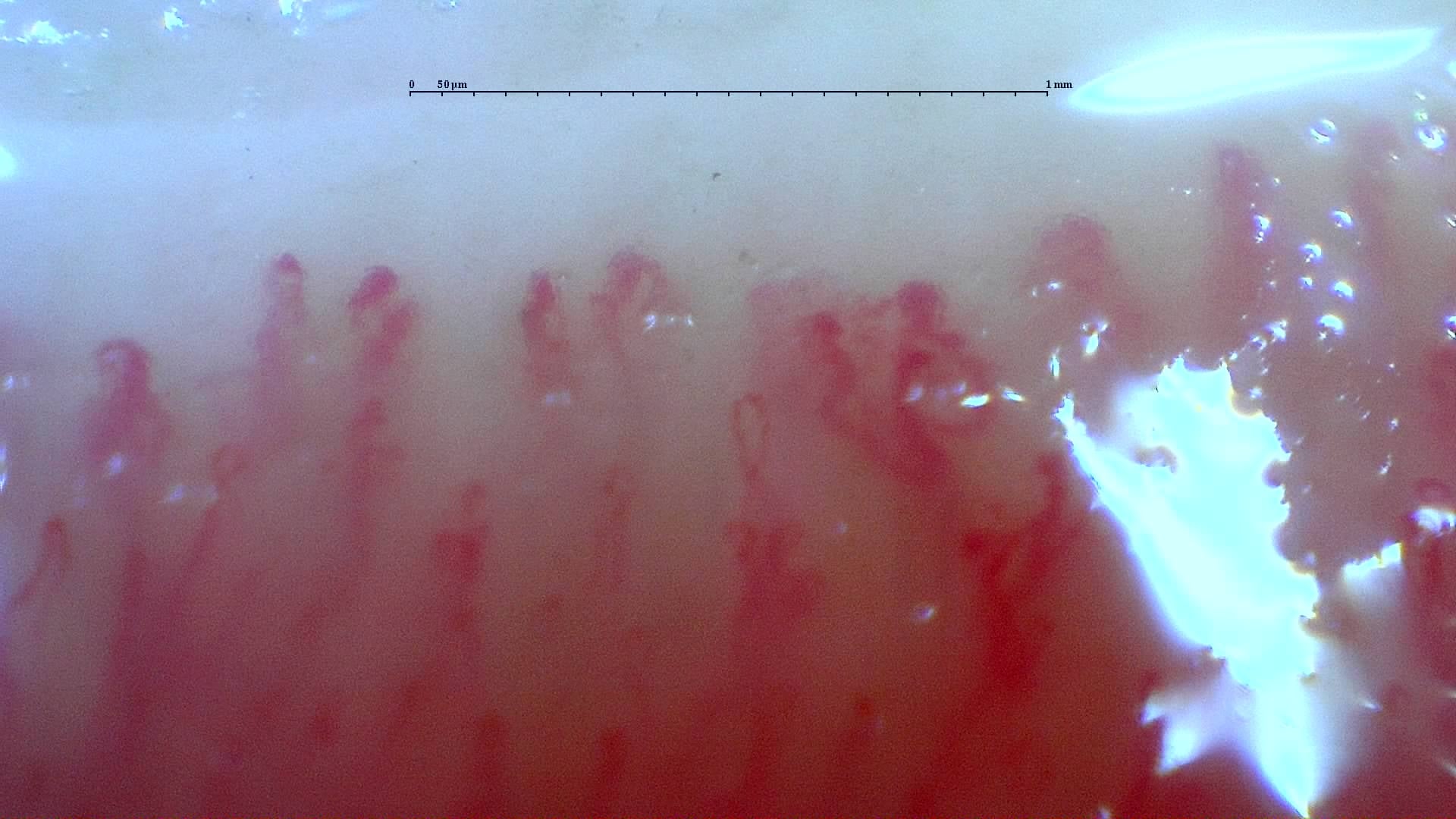
Nailfold capillaroscopy: right little finger, field a.
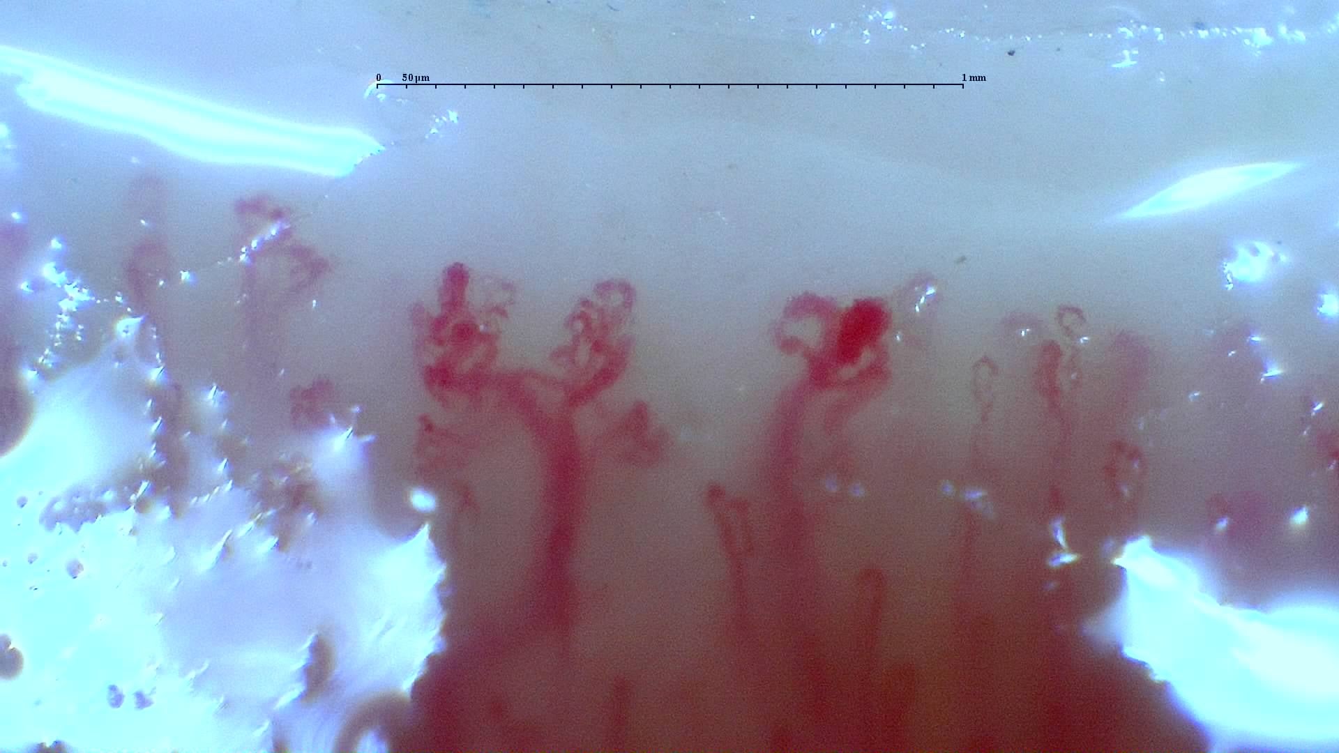
Nailfold capillaroscopy: right little finger, field b.
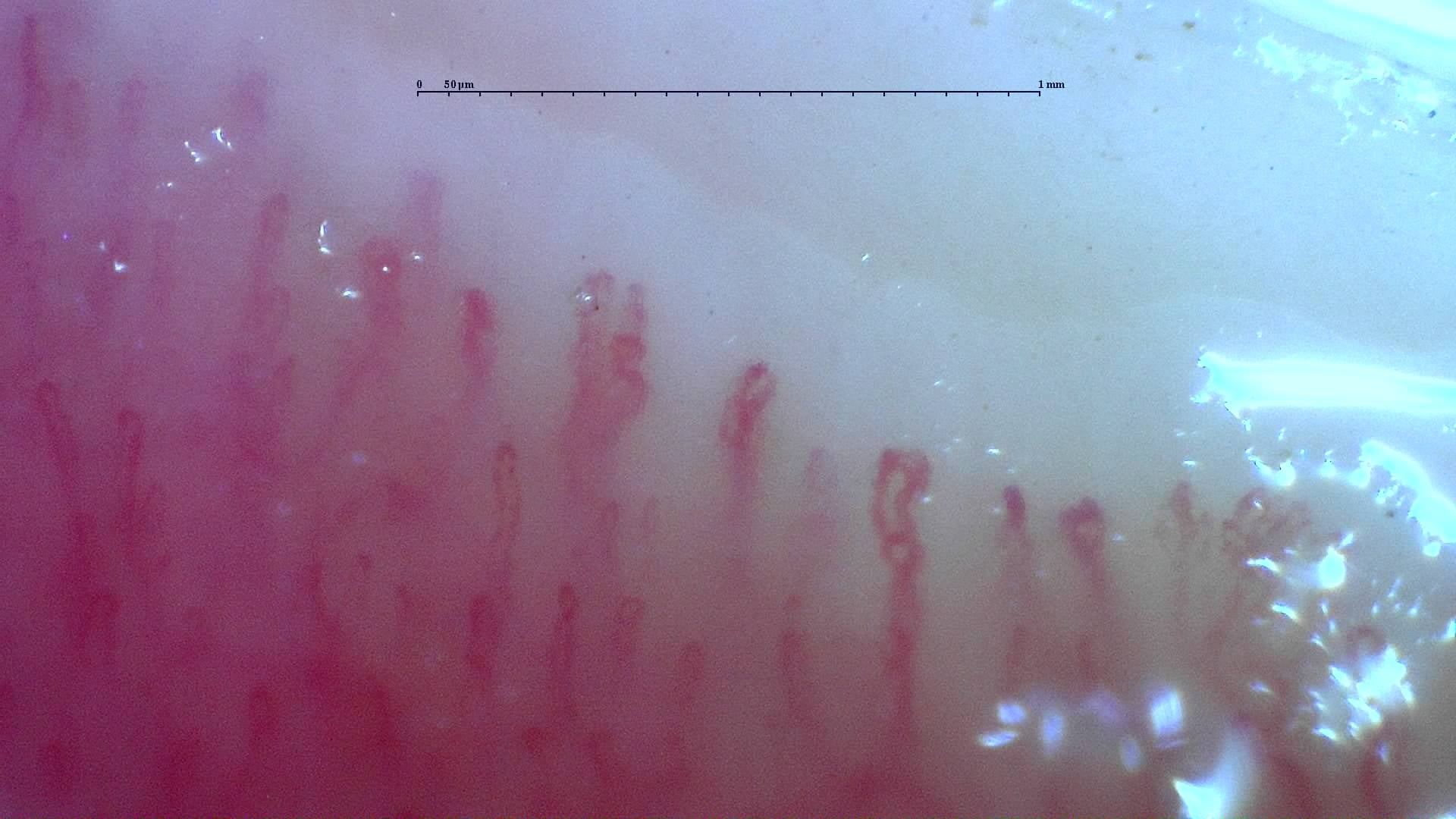
Nailfold capillaroscopy: right little finger, field d.
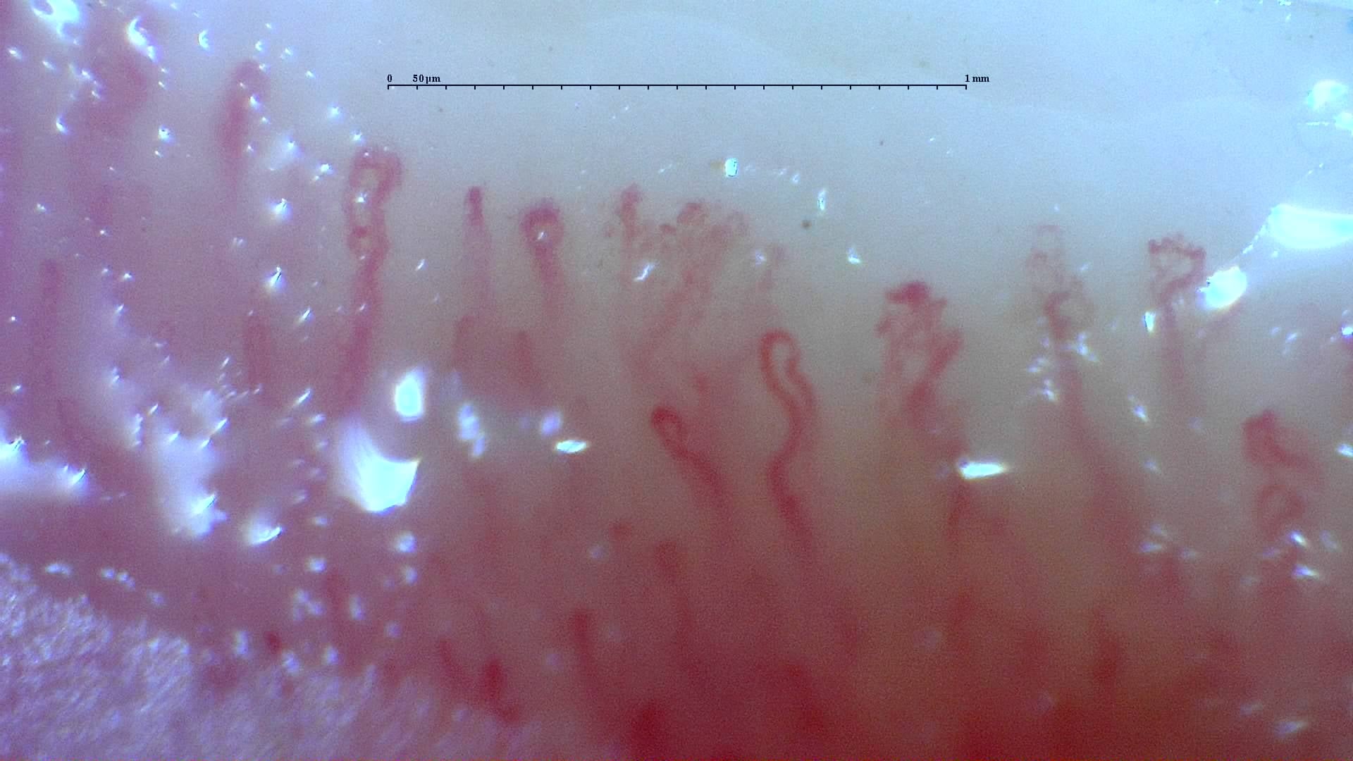
Nailfold capillaroscopy: right little finger, field c.