Interactive viewer
Anticentromere-Positive Systemic Sclerosis with Inter-Digit Pattern Heterogeneity
Images & measurements30The study, field by field
Images & measurements
Explore the fields from each finger and open any image for a closer look. Measurements describe the complete examination at each visit.
Contributed by Miguel Antonio Mesa Navas for educational publication on Capillaroscopy.com. For reuse permissions, contact the rights holder.
Anticentromere-Positive Systemic Sclerosis with Inter-Digit Pattern Heterogeneity
The study is notable for pronounced inter-digit and inter-field heterogeneity. L4C shows marked capillary loss (desertification), whereas L4A has relatively preserved, normal-appearing architecture. R2C and L3D show giant capillaries within a disorganized architecture. R4A demonstrates late-stage revascularization or neoangiogenesis—a morphology the contributor describes as a "post-late" pattern—while R5A shows transitional active-to-late features. The complete study is classified as a late scleroderma pattern, with a mean capillary density of 5.63/mm, enlarged capillaries in 43.5%, and giant capillaries in 2.5%. Image quality is variable; several fields are technically limited and should not be interpreted in isolation.
Recorded measurements
- Capillary density
- 5.63capillaries/mm
- Enlarged capillaries
- 43.53%
- Giant capillaries
- 2.52%
- Hemorrhages
- 0.04/mm
Values from the saved analysis: density and hemorrhages per total analyzed length; percentages of the total capillary count. These are not measurements of an individual image and may differ from averages across fields.
Left index finger
4 images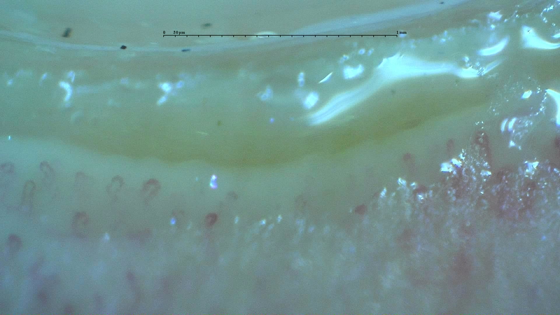
Nailfold capillaroscopy: left index finger, field a.
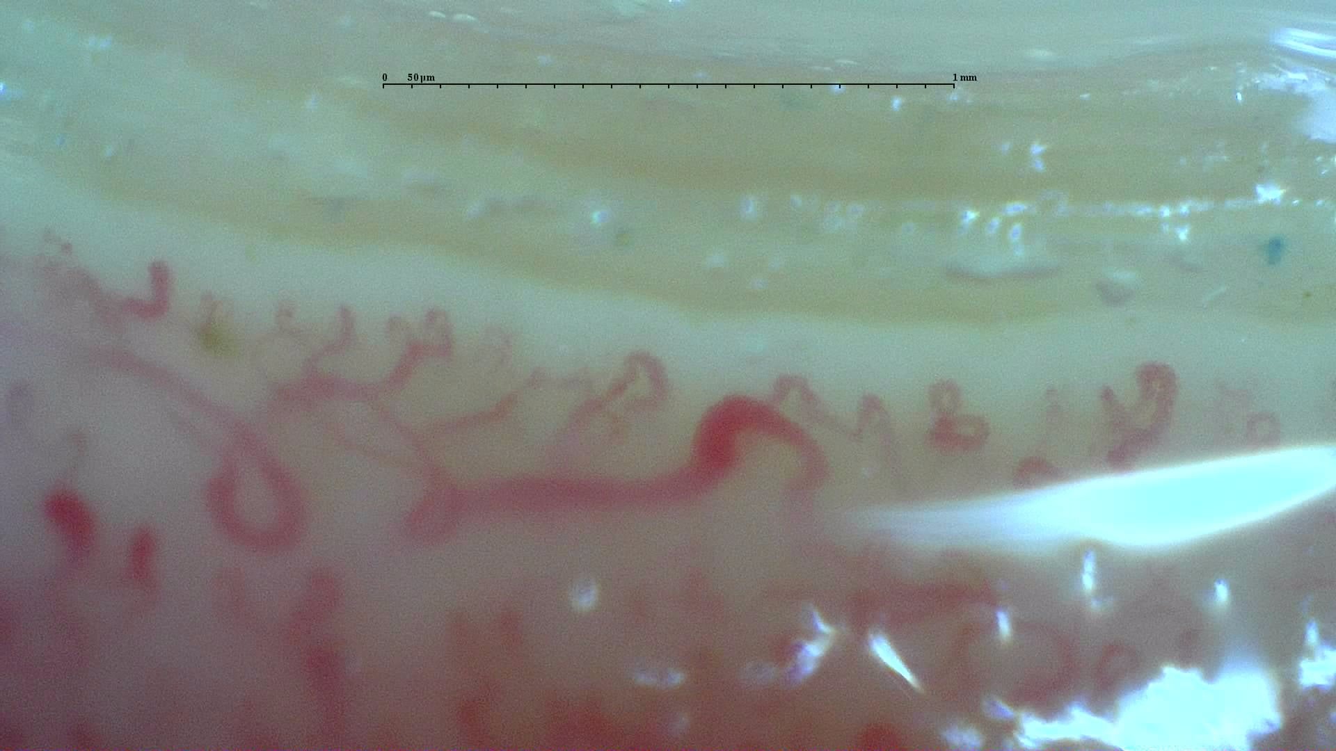
Nailfold capillaroscopy: left index finger, field b.
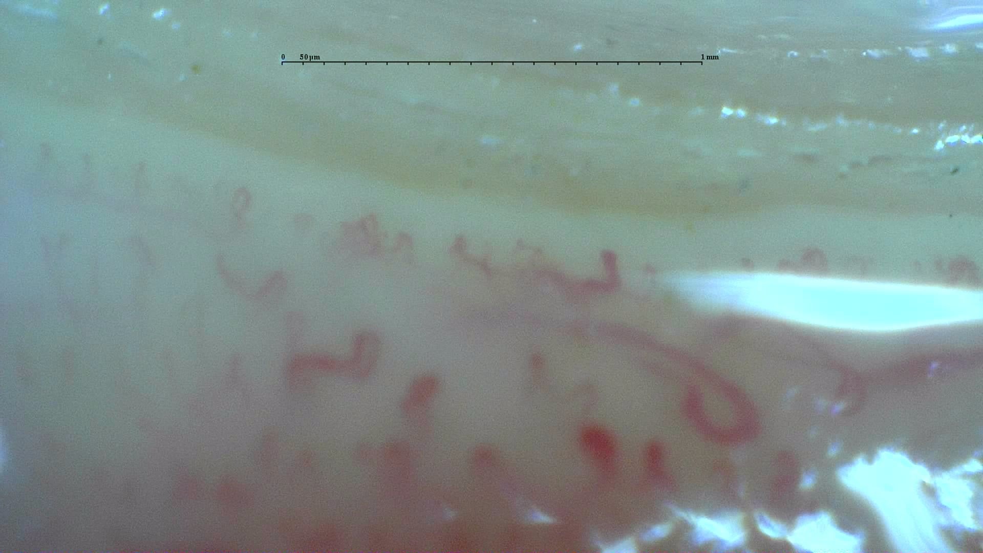
Nailfold capillaroscopy: left index finger, field c.
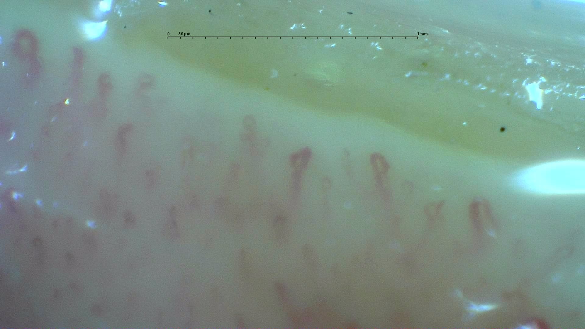
Nailfold capillaroscopy: left index finger, field d.
Left middle finger
4 images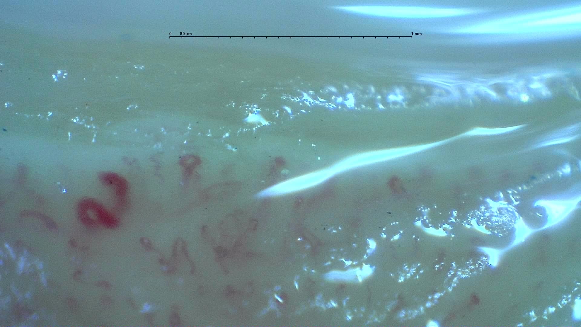
Nailfold capillaroscopy: left middle finger, field a.
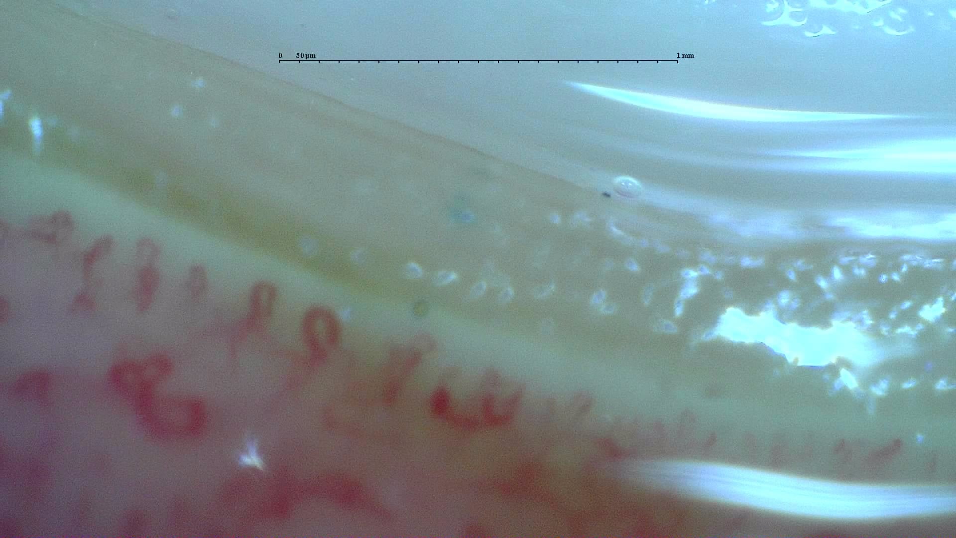
Nailfold capillaroscopy: left middle finger, field b.
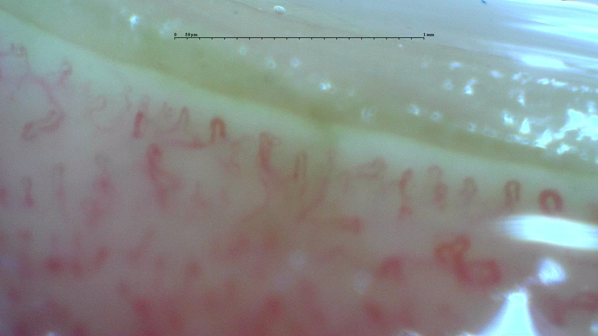
Nailfold capillaroscopy: left middle finger, field c.
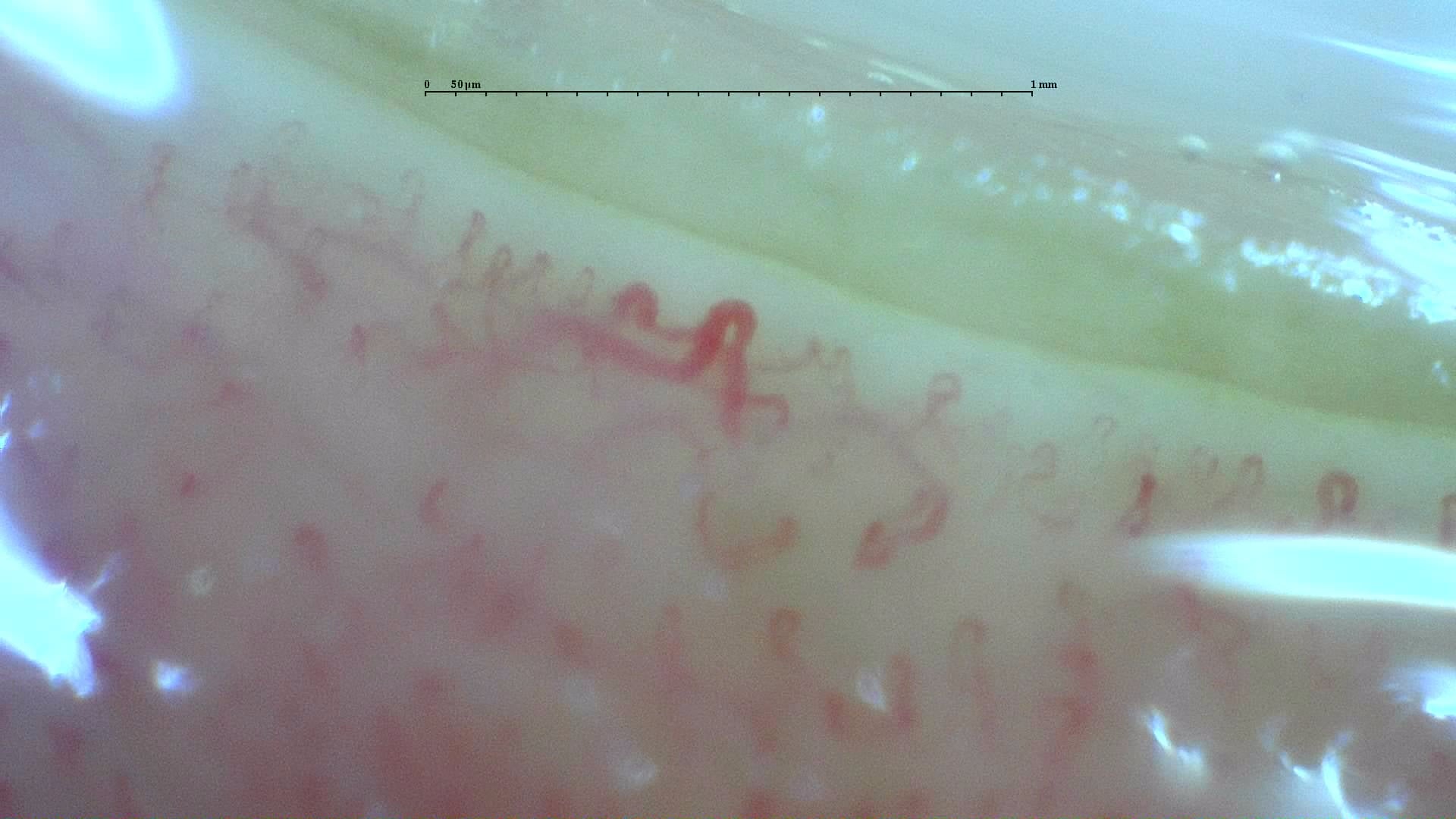
Nailfold capillaroscopy: left middle finger, field d.
Left ring finger
3 images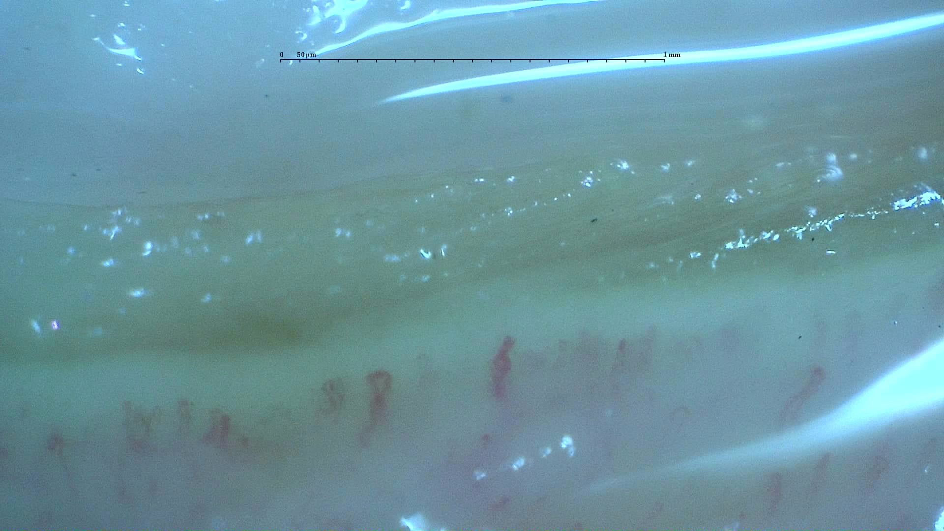
Nailfold capillaroscopy: left ring finger, field a.
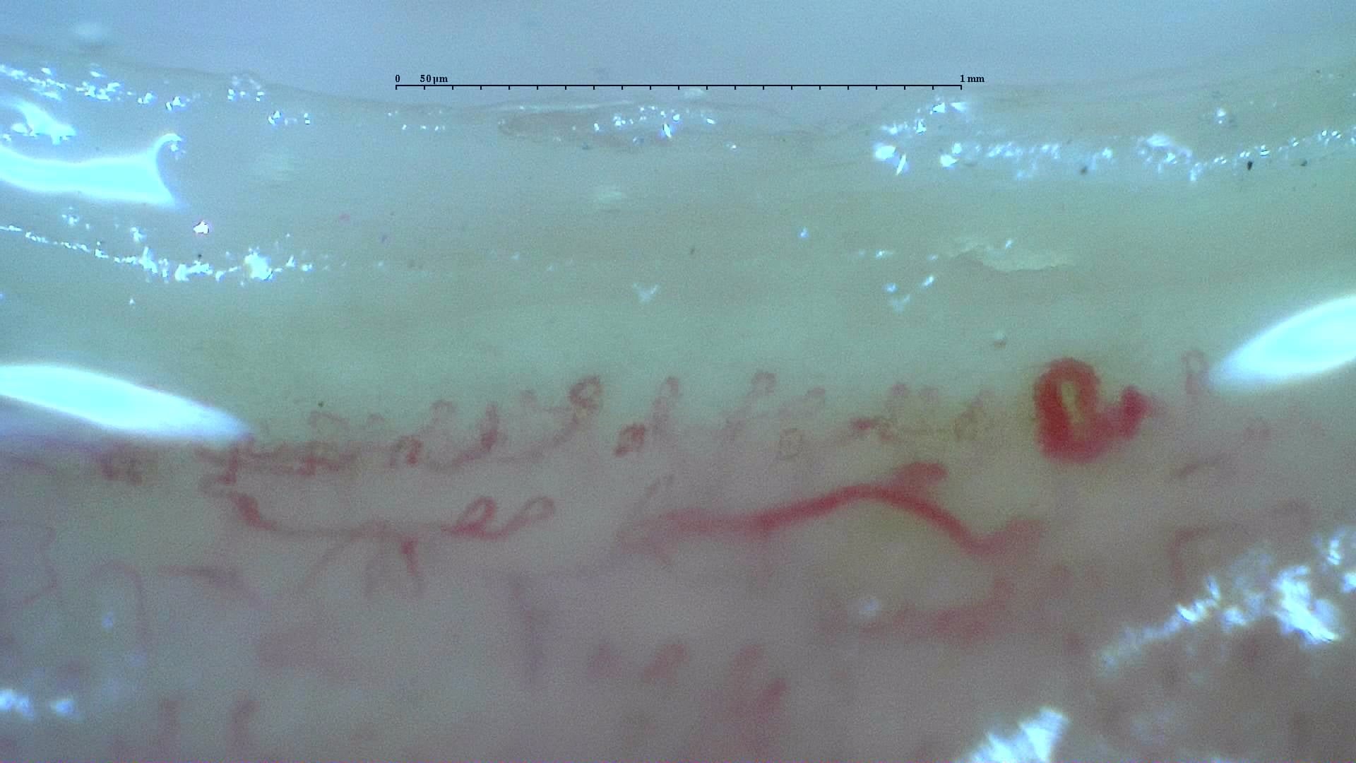
Nailfold capillaroscopy: left ring finger, field b.
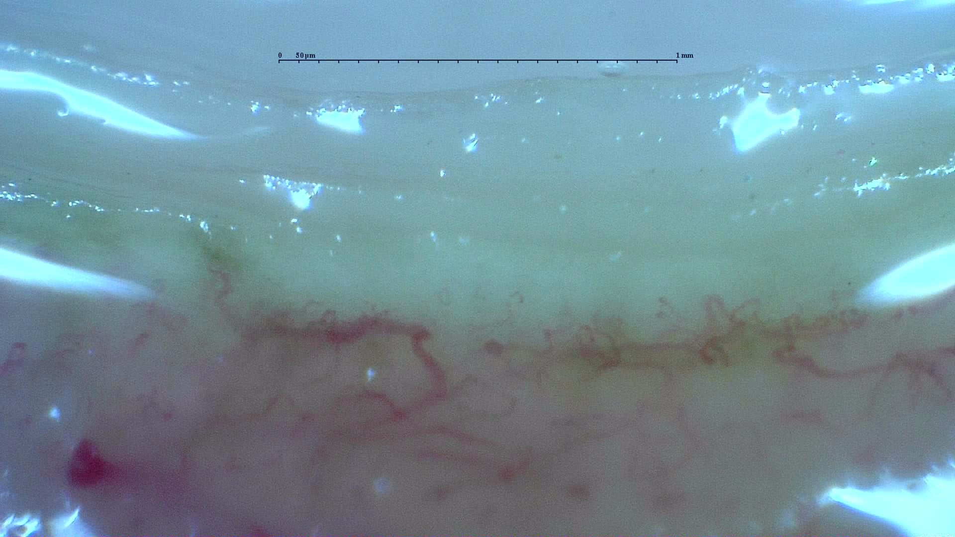
Nailfold capillaroscopy: left ring finger, field c.
Left little finger
3 images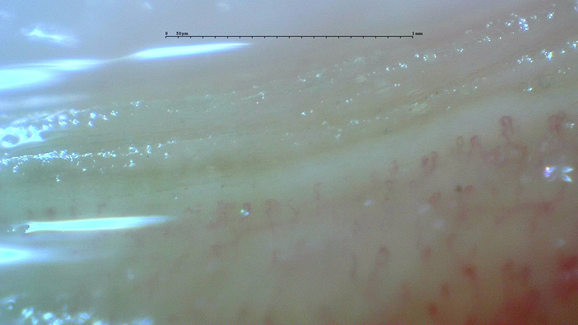
Nailfold capillaroscopy: left little finger, field a.
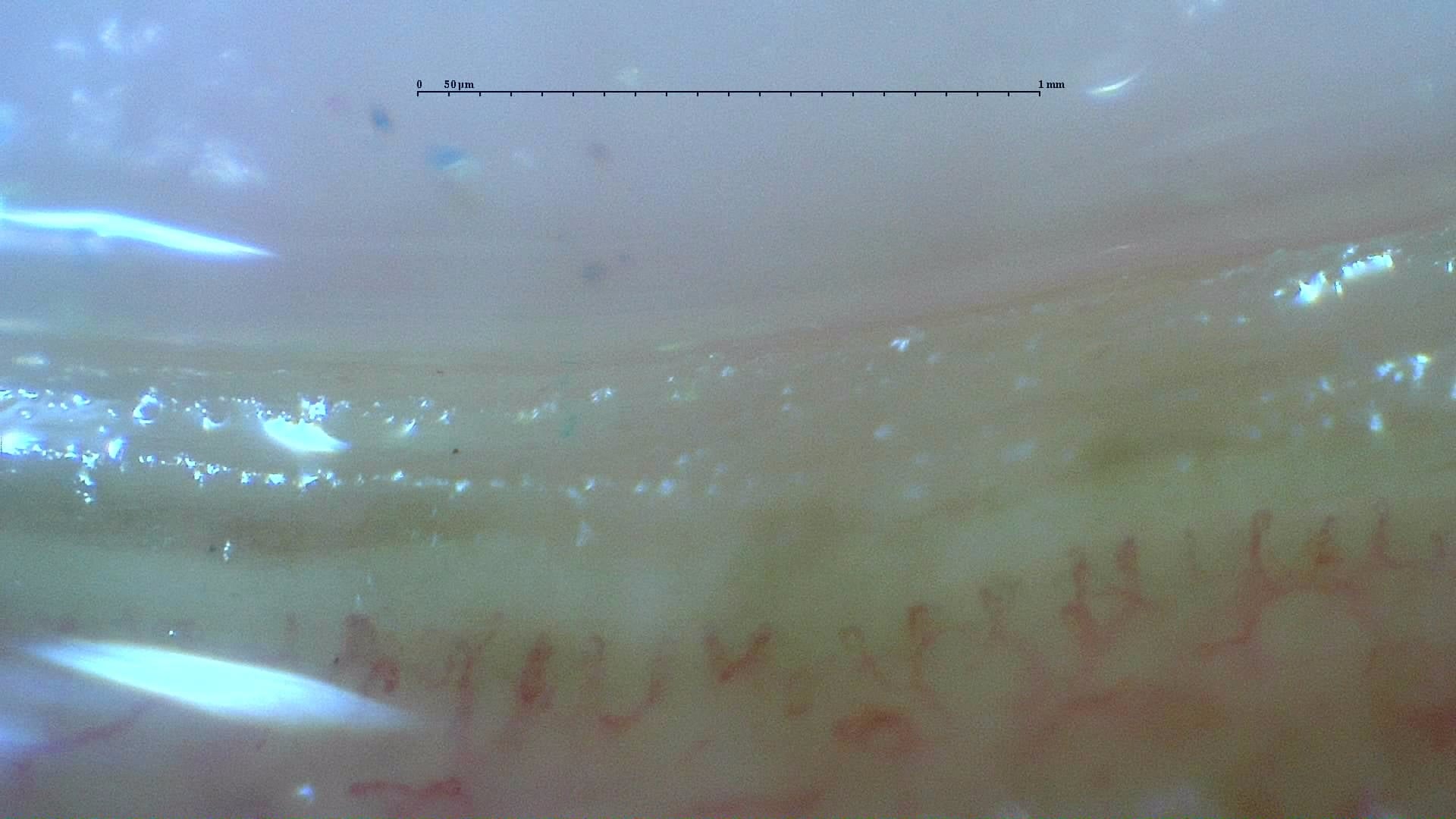
Nailfold capillaroscopy: left little finger, field b.
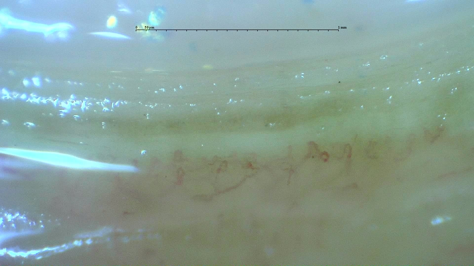
Nailfold capillaroscopy: left little finger, field c.
Right index finger
4 images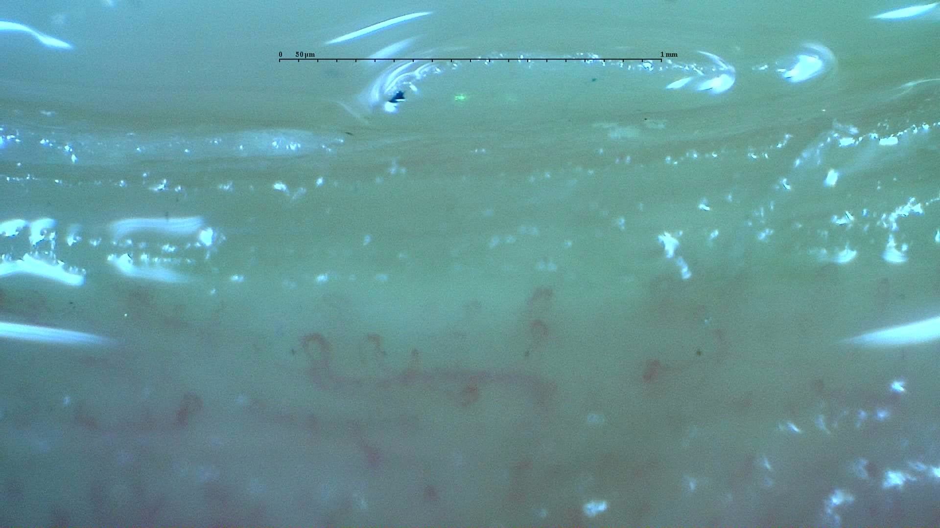
Nailfold capillaroscopy: right index finger, field a.
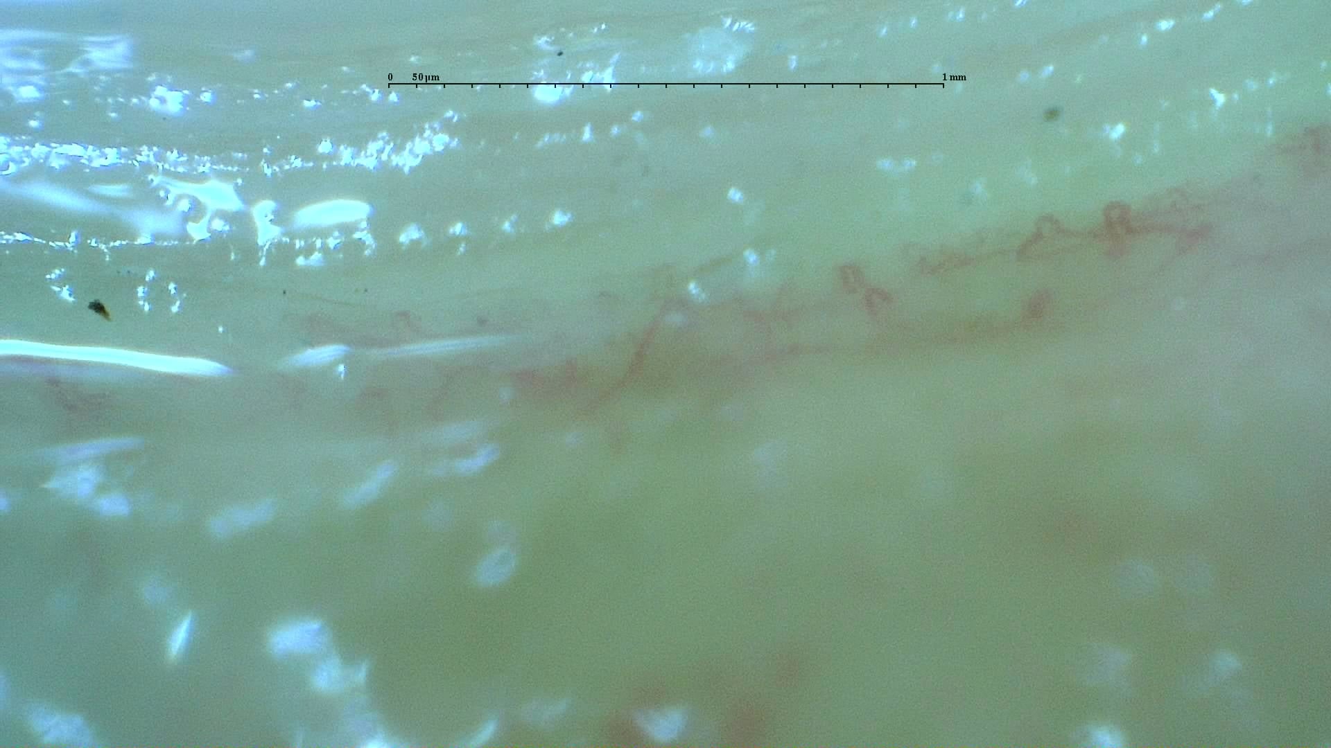
Nailfold capillaroscopy: right index finger, field b.
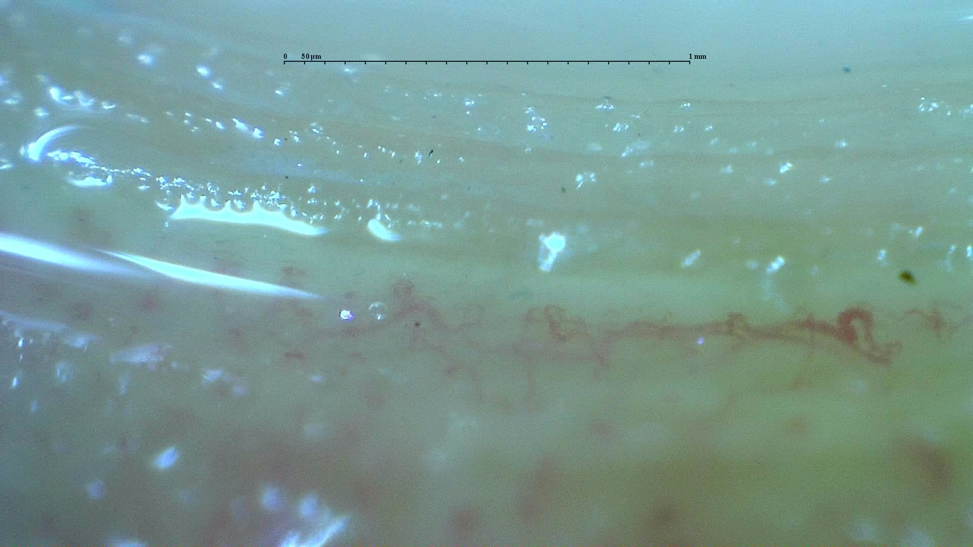
Nailfold capillaroscopy: right index finger, field c.
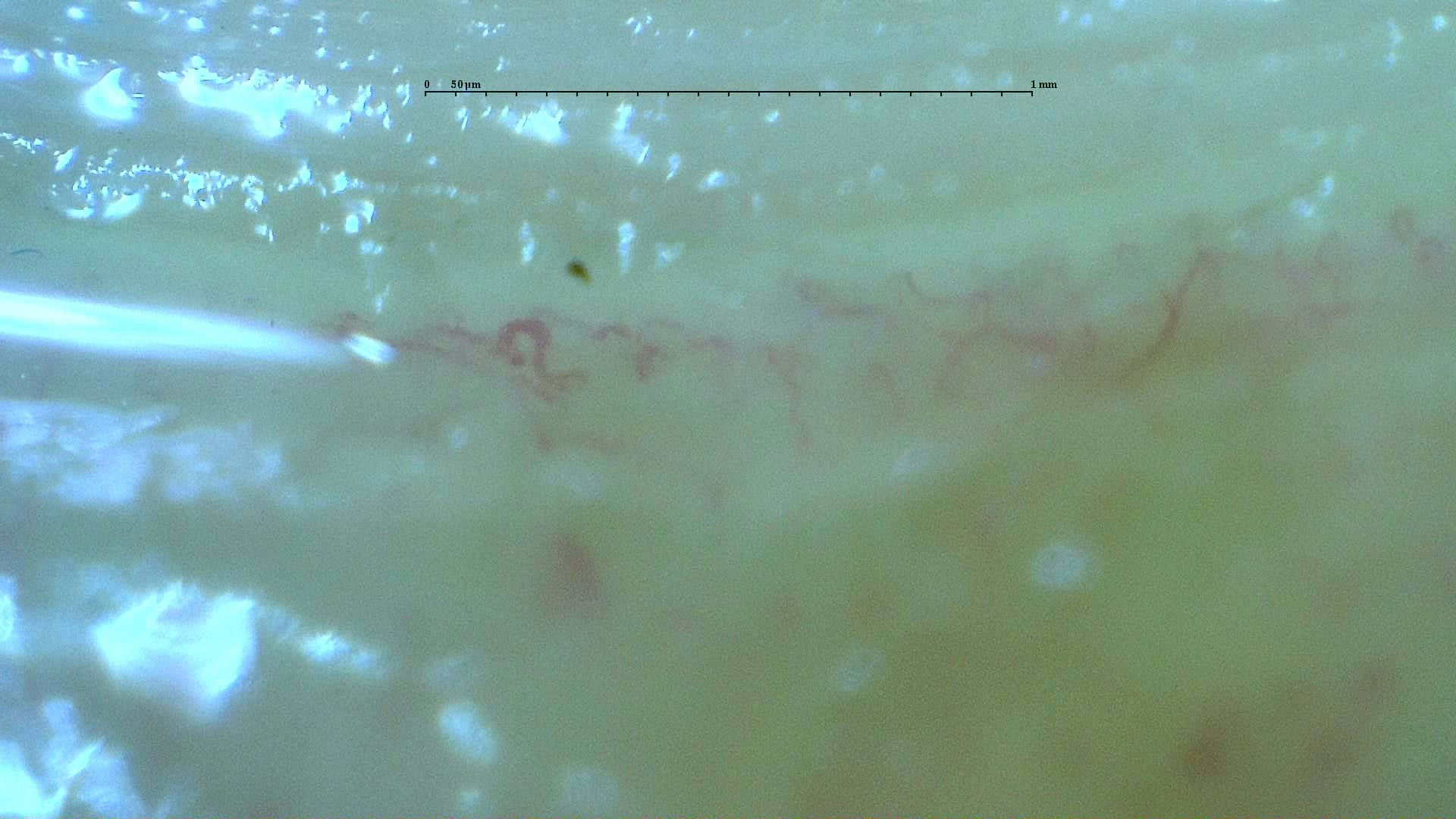
Nailfold capillaroscopy: right index finger, field d.
Right middle finger
4 images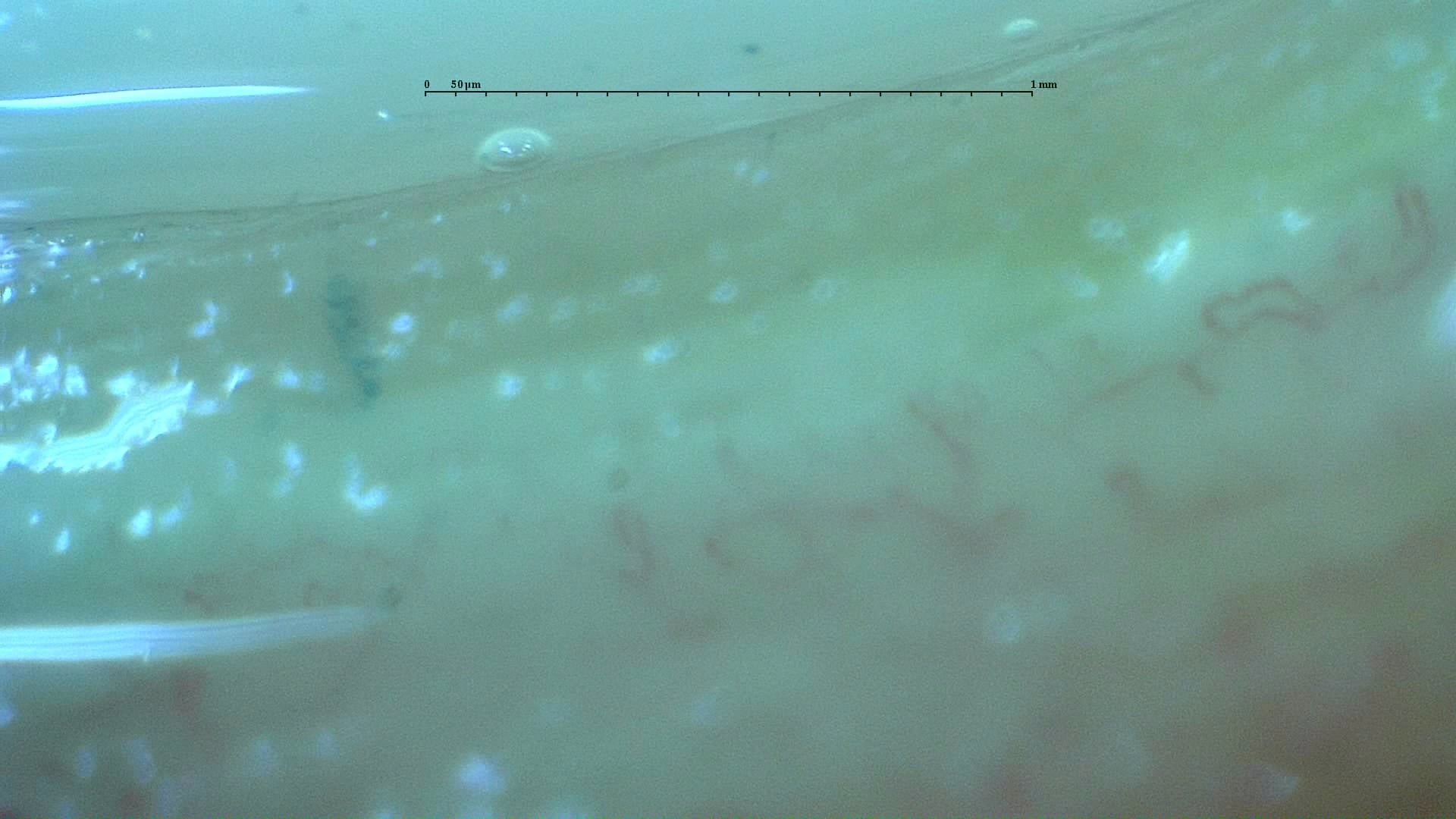
Nailfold capillaroscopy: right middle finger, field a.
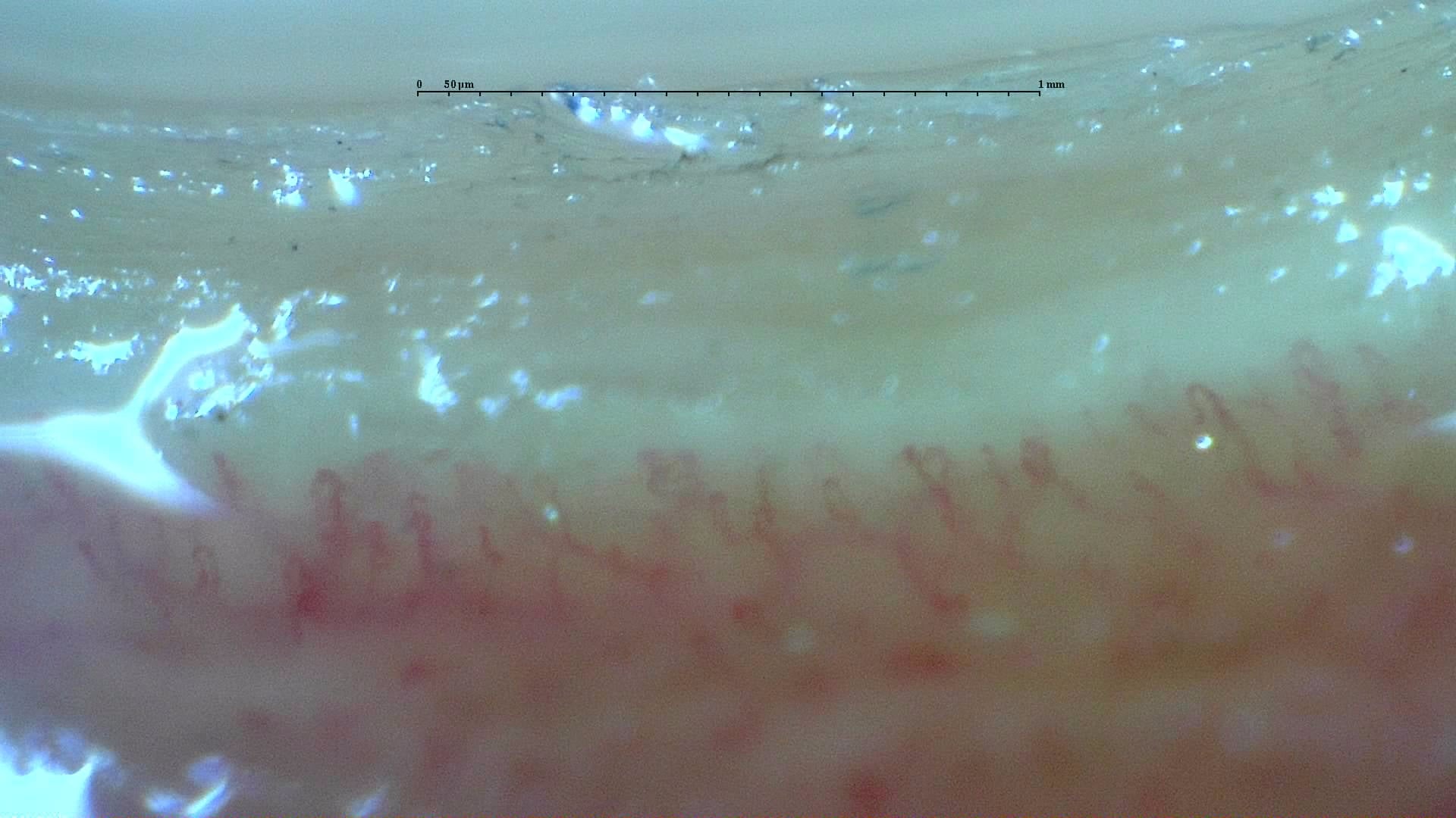
Nailfold capillaroscopy: right middle finger, field b.
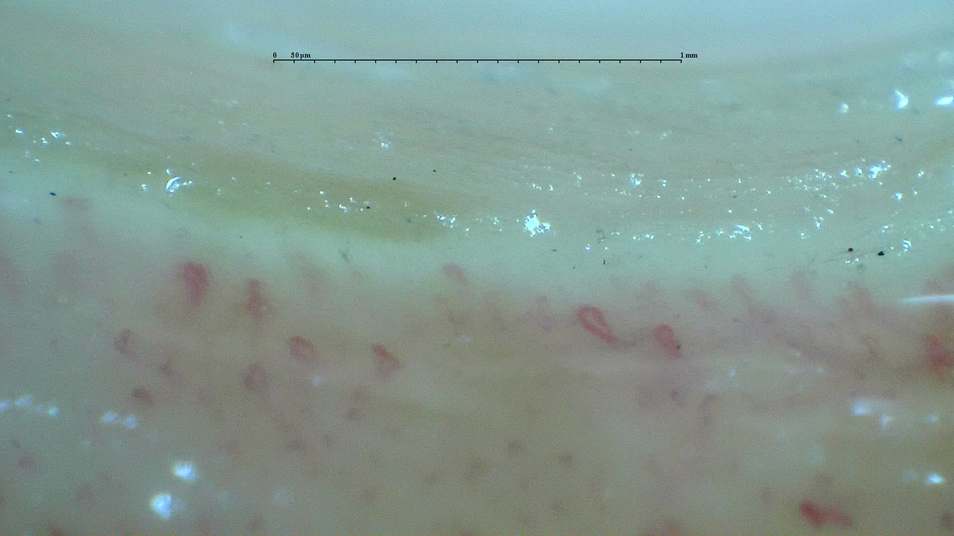
Nailfold capillaroscopy: right middle finger, field c.
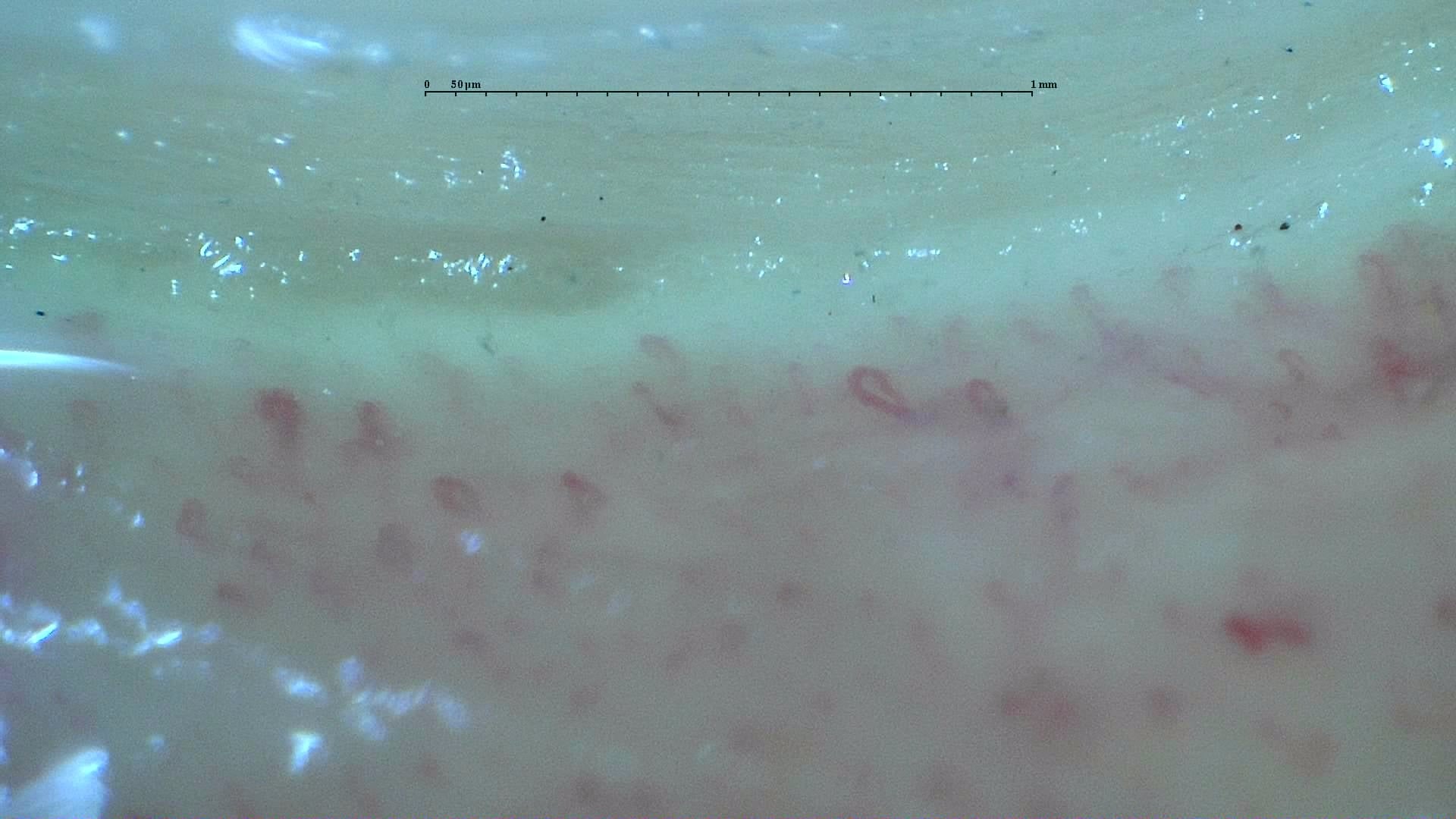
Nailfold capillaroscopy: right middle finger, field d.
Right ring finger
4 images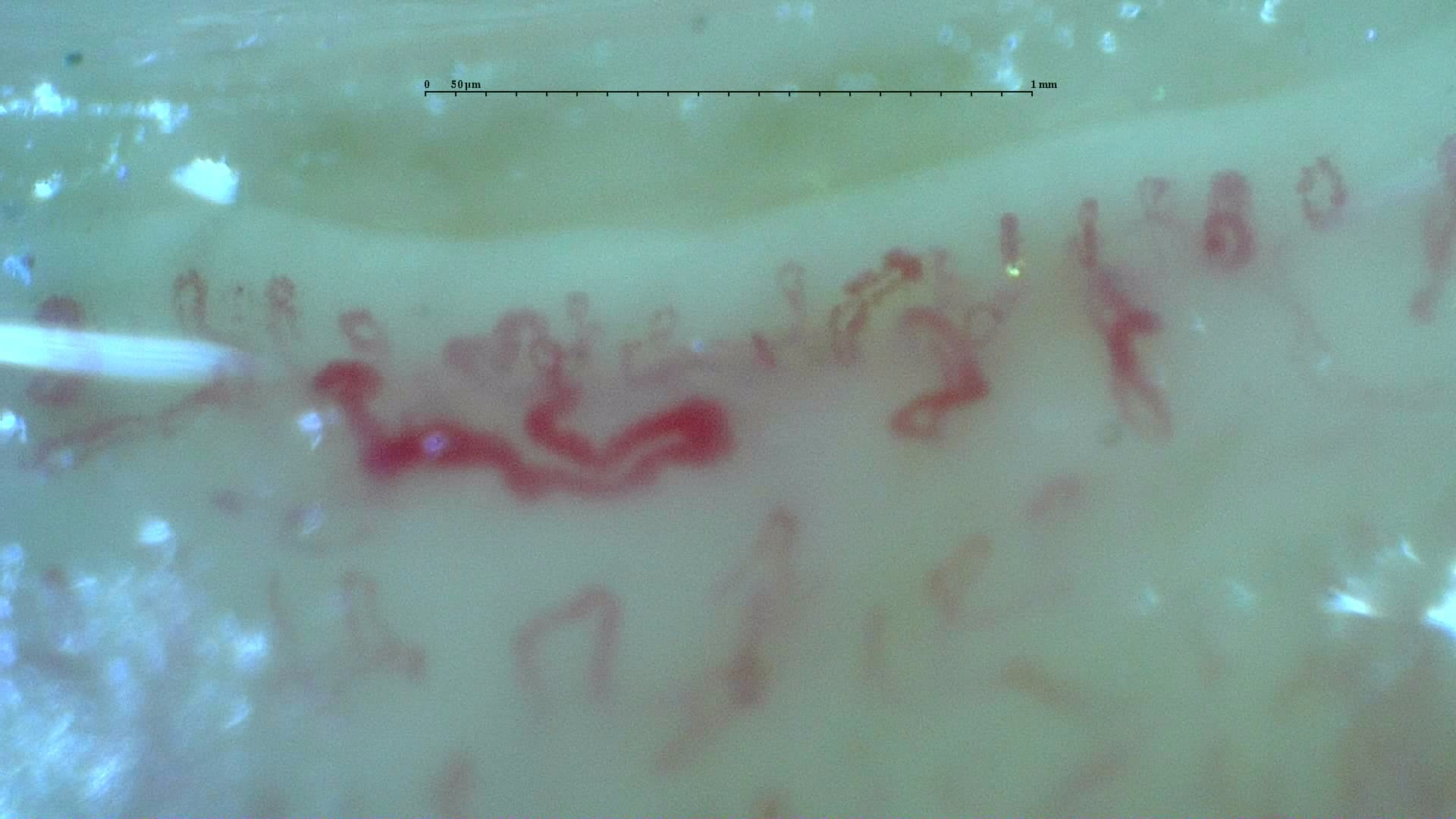
Nailfold capillaroscopy: right ring finger, field a.
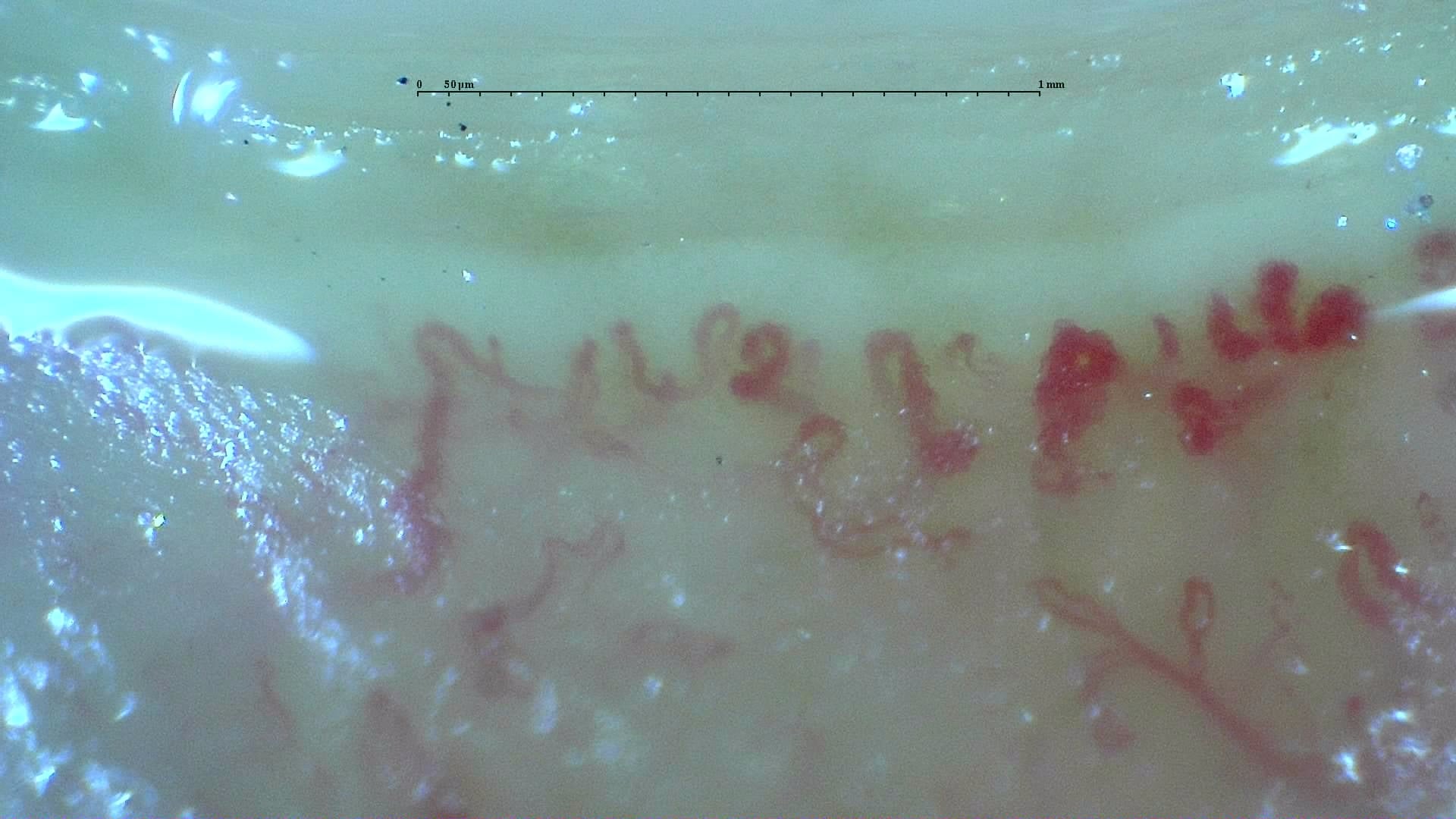
Nailfold capillaroscopy: right ring finger, field b.
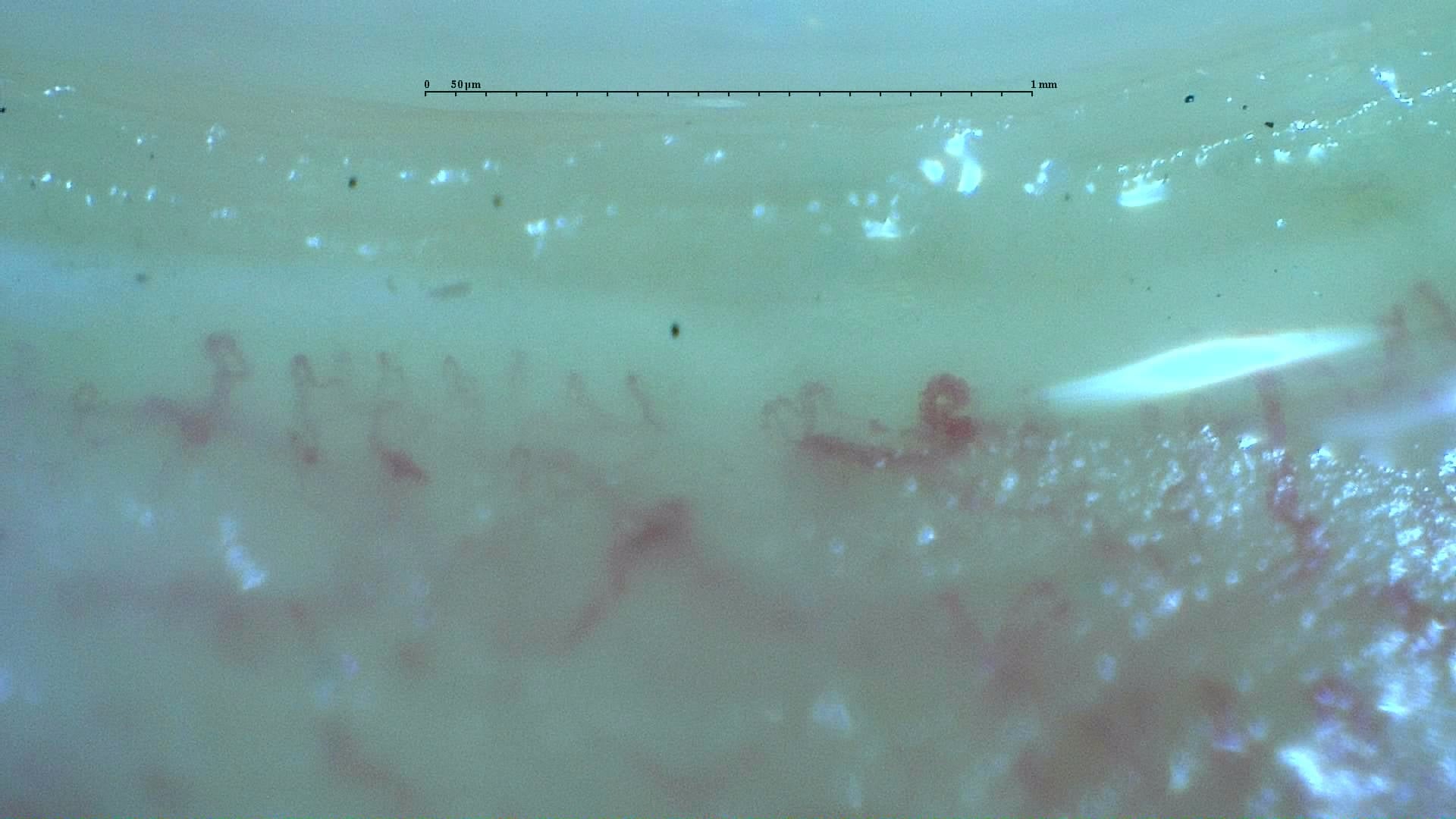
Nailfold capillaroscopy: right ring finger, field c.
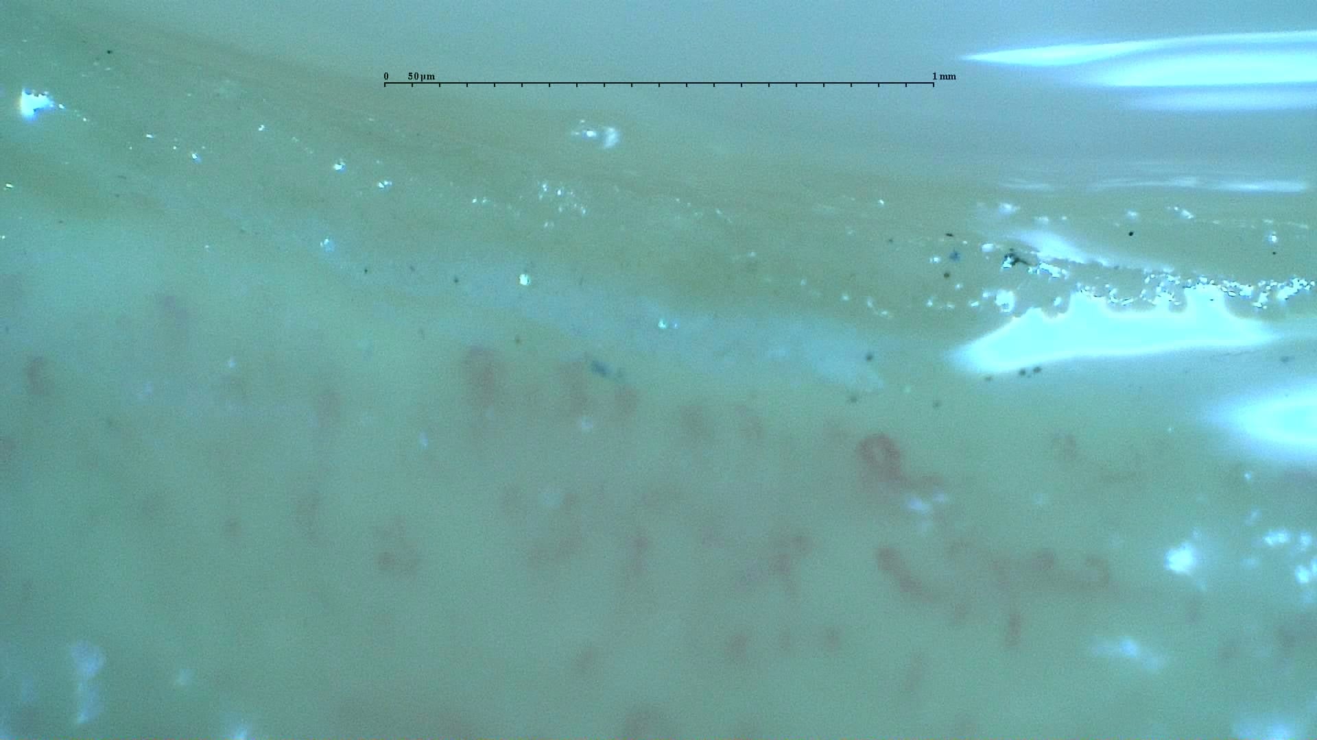
Nailfold capillaroscopy: right ring finger, field d.
Right little finger
4 images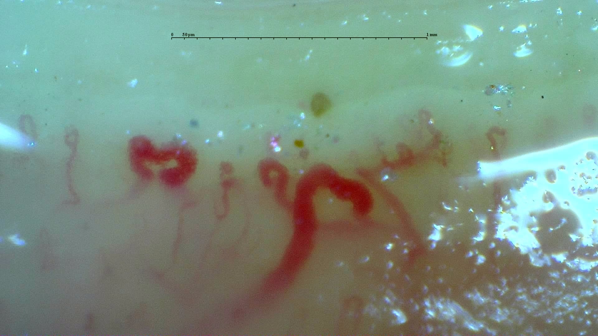
A disorganized field with enlarged loops and surface reflections from the inter-digit heterogeneity case. Other fields differ markedly; inspect the full study and its image-quality caveats.
Read the atlas commentary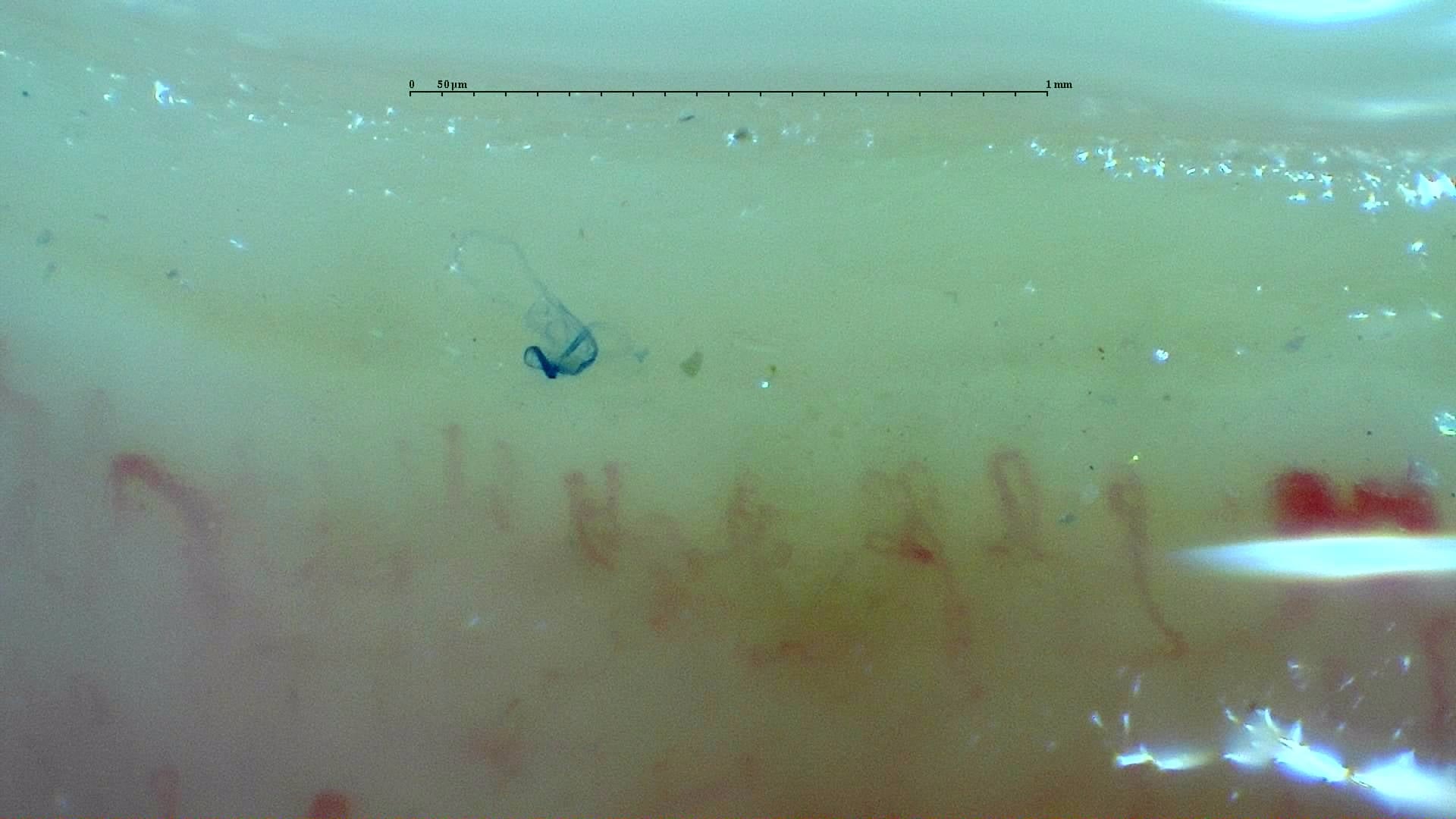
Nailfold capillaroscopy: right little finger, field b.
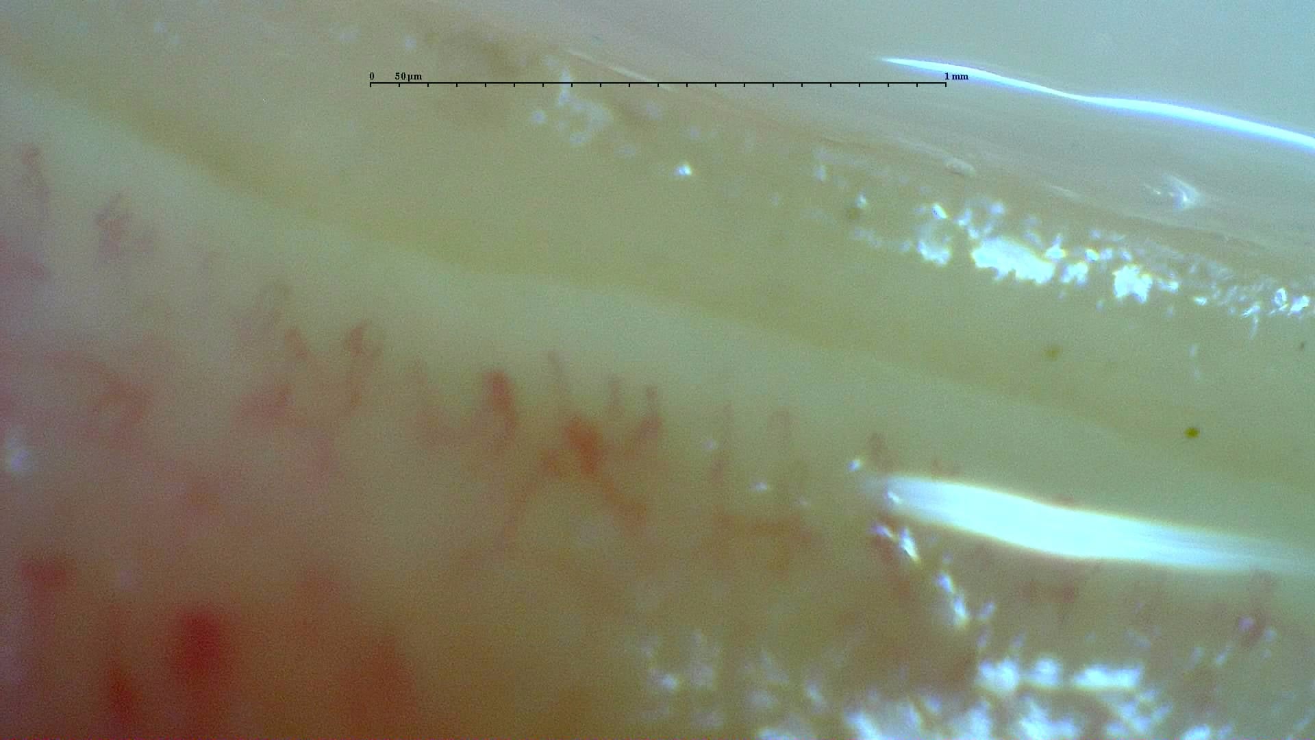
Nailfold capillaroscopy: right little finger, field c.
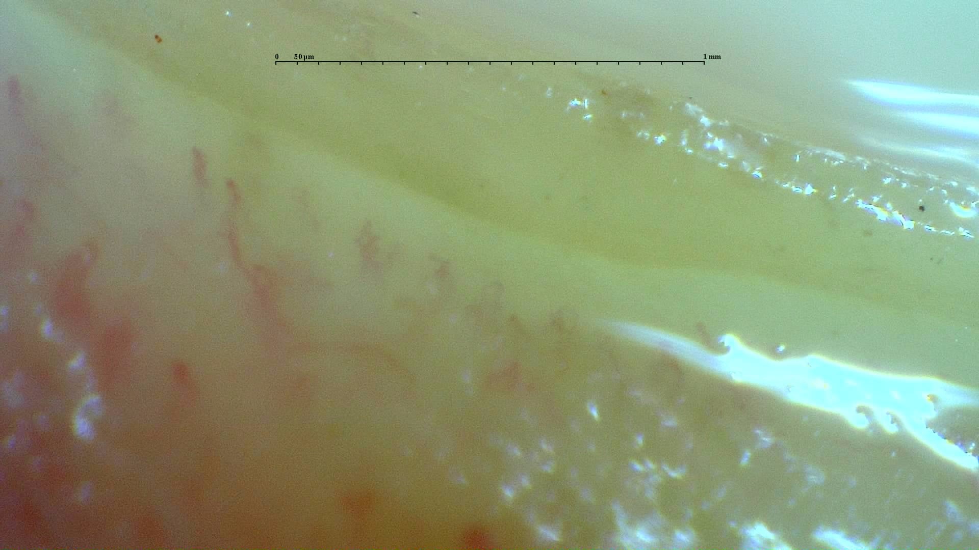
Nailfold capillaroscopy: right little finger, field d.